

The cow skeleton is the hard framework of its body and supports the soft internal organs and structures. This article will help you to learn the detail anatomical features of the cow’s skeleton with diagrams.
Quick answer: The cow skeleton is a hard framework and consists of bones, cartilages, and ligaments. It is responsible for the maintenance of shape, flexibility, and locomotion of the cow’s body.
Here, I will focus on the description of the main component (bones) of the cow’s skeleton in detail. Thus, you will know and identify all the bones from different regions of the cow’s skeleton perfectly.
Cow skeleton
Having good knowledge of the animal’s skeleton is the key requirement for future deep learning of veterinary anatomy. Most of the bones from the cow skeleton represent the typical osteological features compared to other animals.
Thus, you need to learn the bones from the cow’s skeleton carefully, so that you may compare the osteological features with other animal species while reading special anatomy.
First, let’s know what a skeleton is and what the types of skeletons are in animals, or cows.
What is the skeleton in a cow?
Definition: Skeleton in a cow is the hardest structure that provides the supporting framework of its body and protects internal soft tissues. It also provides a firm base to which the muscles are attached and helps in locomotion.
Thus, the external structure and appearance of animals are provided by the skeleton. The cow’s skeleton is made of three components –
- Bones: bones are the principal component of the skeleton,
- Cartilages: they are the specialized connective tissue structures that connect to the ends of the bones.
- Ligaments: They are the bands or membranes of fibrous connective tissue that bind bones together.
Figure 1 shows the bones, cartilages, and ligaments cow’s skeleton. It also represents how it protects the visceral organs of the body.

Bones from different parts of the cow’s skeleton play an important part in the function of the skeletal system. Again, they have a specific descriptive name and function that contributes to the movement of the body.
For description purposes, a cow’s skeleton is mainly divided into two parts –
- Axial skeleton: This part of the skeleton is arranged along the long axis of the cow’s body. It includes the bones of the skull, vertebral column, ribs, and sternum.
- Appendicular skeleton: This part of the skeleton is arranged in the vertical position of the body. It includes the bones of the forelimb and hindlimb of the cow or other animals.
Here, Figure 2 shows the axial and appendicular skeletons of the cow. It also identifies the bones from these two parts of the skeleton.

Visceral or splanchnic bones in the skeleton
In some of the organs of a few animals and birds, there are visceral or splanchnic bones that are embedded in the parenchyma of that specific organ. They are the modified musculature of that specific organ and have no connection to the skeleton.
Bones identification from a cow skeleton
It is better to identify all of the bones from the cow’s skeleton according to the axial and appendicular skeleton.
From the axial skeleton, you will identify the following –
- Bones of the cow skull: facial and cranial bones,
- Vertebrae bone from the vertebral column of the cow: cervical, thoracic, lumbar, sacral, and coccygeal vertebrae,
- Ribs and sternum bones: 13 pairs of ribs and 1 sternum,
From the appendicular skeleton, you will identify the following –
- Bones of the forelimb of a cow: first bone -scapula, humerus, radius and ulna, carpals, metacarpals, sesamoids, and phalanges,
- Bones from the hindlimb of a cow: hip bones, femur, patella, tibia and fibula, tarsals, metatarsals, sesamoids, and phalanges.
Now, let’s know the names of the bones of the skull, vertebrae, and limbs from the cow’s skeleton. Figure 3 shows the names of the different bones from the cow’s skull, vertebrae, and limbs (but you will know more details of these bones).

How many bones are in the cow’s skeleton?
Bone count in a cow’s skeleton: There at 204-206 bones in the cow’s skeleton. The name and number of bones are shown in Table 1 –
| Name of bones | Number of bones |
| Skull bones Facial bones Cranial bones Hyoid bones Mandible bones | Total: 32 17 11 02 02 |
| Bones in the vertebral column Cervical vertebra Thoracic vertebra Lumbar vertebra Sacral vertebra Coccygeal vertebra | Total: 49 – 51 07 13 06 05 18-20 |
| Ribs and sternum of the cow Sternal ribs (8 pairs in each side) Asternal ribs (8 pairs in each side) Floating ribs Sternum bone | Total: 27 8×2 = 16 5×2 = 10 0 01 |
| Bones from the forelimb (right+left) Scapula (1 in each side) Humerus (1 in each side) Radius and ulna (2 in each side) Carpal bones (6 in each side) Metacarpal bones Large metacarpal (1 in each side) Small metacarpal (1 in each side) Proximal sesamoids(4 in each side) Phalanges (3 in each digit) Distal sesamoid (1 in each digit) | Total: 48 02 02 2×2= 4 6×2 = 12 1×2= 2 1×2 = 2 4×2 = 8 3×4 = 12 1×4 = 4 |
| Bones of the hindlimb (right+left) Hip bones (1 in each side) Femur bone (1 in each side) Patella (1 in each side) Tibia and fibula (2 in each side) Tarsal bones (5 in each side) Metatarsal bones (1 in each side) Proximal sesamoids (2 in each side) Phalanges (3 in each digit) Distal sesamoids (1 in each digit) | Total: 46 2 2 2 4 10 2 8 12 4 |
| Visceral bones | 2 |
| Total bones in cow | 204 – 206 |
Cow skull bones identification
The skull of a cow consists of facial and cranial bones. Some of the facial and cranial bones of the cow’s skull are paired, and some are unpaired.
Here, I will help you to identify the cranial and facial bones only from the cow skull. Table 2 shows the facial and cranial bones from the cow skull with their number.
| Facial bones of the cow skull | Cranial bones of the cow skull |
| Lacrimal bone (paired) Zygomatic / malar (paired) Maxilla bone (paired) Pre-maxilla (paired) Palatine (paired) Nasal (paired) Pterygoid (paired) Turbinate bone (paired) Vomer (single) | Occipital (single) Parietal (paired) Interparietal (paired) Frontal (paired) Temporal bone (paired) Sphenoid bone (single) Ethmoid bone (single) |
Most of the facial and cranial bones from the cow skull are identified in Figure 4. But it is better to identify these bones randomly from different views.

For example, you may identify the bones from the dorsal view first. Then you may identify the cow’s skull bones from the lateral view. Finally, you may identify the bones from the ventral view of the cow’s skull.
Cow’s hyoid bone identification
From the cow’s hyoid bone, you might identify the following parts –
- Stylohyoid bone (Great cornu),
- Ceratohyoid bone (middle cornu),
- Lingual process of the hyoid bone,
- Basihyoid bone (body of hyoid), and
- Thyrohyoid bone (thyroid cornu),
Here, Figure 5 shows the different segments or parts of the cow’s hyoid bone.

Cow mandible identification
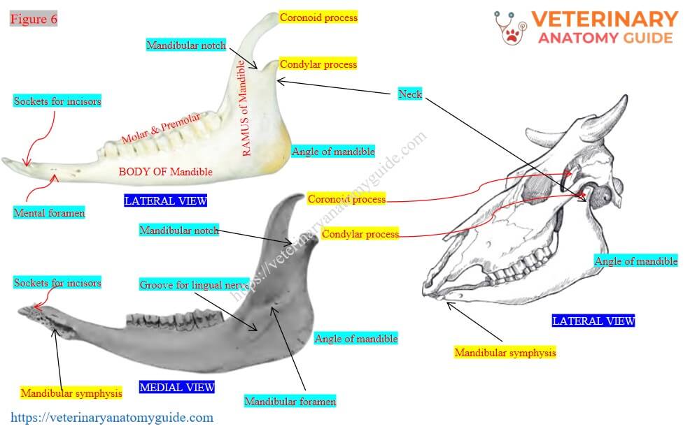
The mandible of the cow consists of a horizontal body and a vertical ramus. The two halves of the mandible fuse incompletely at the midline of the body and form a single mandible.
From the body of the cow mandible, you might identify the following osteological features –
- 2 surfaces, 2 borders, and 2 extremities,
- 8 alveolar sockets for incisor teeth (anteriorly),
- 6 alveolar sockets for cheek teeth (on the dorsal border of the body),
- Mental foramen at the anterior aspect of the lateral surface,
- The mandibular canal passes through the body to the mandibular foramen.
From the vertical ramus of the cow’s mandible, you might identify the following osteological features –
- 2 surfaces, 2 borders, and 2 extremities,
- Condylar and coronoid processes at the proximal extremity of the ramus,
- Mandibular notch (MN) between the coronoid and condylar processes,
- Neck of the mandible,
- Mandibular foramen and a groove for the lingual nerve on the medial surface, and
- Angle of mandible with a faint notch for the maxillary vessel and parotid duct,
Figure 6 shows the different osteological features of the body and ramus of a cow mandible.
Vertebrae identification from the cow skeleton
Vertebrae are the single, unpaired, and irregular bones in the cow’s skeleton. They are articulate together serially and form a long column along the axial skeleton of the cow’s body, which is known as the vertebral column or spine.
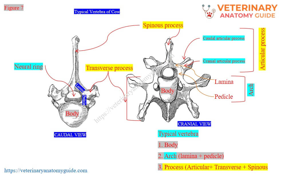
Before going to identifying the osteological features of different vertebrae from the vertebral column, you might know the features of a typical vertebra. A typical vertebrae possess three main parts –
- Body: body is cylindrical and possesses two extremities. Here, the cranial extremity is convex, while the caudal extremity is concave.
- Arch: arch forms the two lateral walls of the vertebral ring and consists of the dorsal lamina and ventral pedicle.
- Processes: there are 3 processes – spinous, transverse, and articular. Here, the spinous process is single, the transverse process is paired and projects outward from the lateral part of the neural ring. Again, the articular process consists of paired cranial and paired caudal segments.
Figure 7 shows the different parts of a typical vertebra of a cow.
Vertebral bones are divided into 5 regions according to their position and possess similar osteological features –
- Cervical vertebrae: 7 in number and have long bodies and short processes. But the first and second cervical are highly modified.
- Thoracic vertebrae: 13 in number and have long spinous processes.
- Lumbar vertebrae: 6 in number and have long, flattened transverse processes.
- Sacral vertebrae: 5 in number and fused to form a single bone called the sacrum in the cow.
- Coccygeal vertebrae: vary in number in cows (18 – 20) and are highly atypical.
Cervical vertebrae of a cow
Among the 7 cervical vertebrae of a cow, the first and second (atlas and axis) are highly modified. Again, the sixth and seventh (6th & 7th) cervical vertebrae of a cow also possess unique features. Thus, the third, fourth, and fifth possess the typical features of a vertebra.
The common features of cervical vertebrae of a cow are (except atlas and axis) –
- Body: long, and ventral crest prominent,
- Arches: well-developed in cervical vertebrae,
- Spinous process: short, tuberous, and inclined forward,
- Transverse process: divides into upper and lower segments,
- Transverse foramen: the base of the transverse process is perforated by the foramen transversarium, and
- Articular process: well-developed in cervical vertebrae,
The following articles might help you to learn the osteological features of different cervical vertebrae of a cow –
| First cervical vertebrae | Cow atlas anatomy |
| Second cervical vertebrae | Cow axis anatomy |
| Sixth and seventh cervical | How will you differentiate the sixth cervical vertebrae from the seventh? |
The exceptional features of cow’s cervical vertebrae: First (atlas) and second (axis) cervical vertebrae of a cow are highly modified in their structures. The sixth cervical vertebrae of a cow have a well-developed spine. Again, the lower part of the transverse process of the cow’s sixth cervical is in the form of a quadrilateral plate.
The seventh cervical vertebrae of a cow possess two caudal facets on its caudal part of its body. These facets are for the articulation with the heads of the first pair of sternal ribs of the cow.
The spinous process is well-developed in the cow’s seventh cervical vertebrae. Again, the foramen transversarium is absent in the cow’s seventh cervical vertebrae.
Figure 8 shows the cow’s cervical vertebrae (from first to seventh).

Cow’s thoracic vertebrae
The thoracic vertebrae of a cow form the roof of the thoracic cavity and cage along with the proximal end of the ribs. You will find 13 thoracic vertebrae in the cow’s vertebral column, and they possess the following common osteological features –
- Body: The body of the cow’s thoracic vertebrae is comparatively short and cylindrical,
- Arches: you will find additional intervertebral foramina at each side of the arches,
- Spinous process: Spinous processes are long and directed backward in the cow.
- Transverse process: they are short in the cow and undivided.
- Articular process: They are not well-developed in the thoracic vertebrae. The cranial articular facets of the cow’s thoracic vertebrae are in the form of small oval articular facets.
How is the intervertebral foramen formed?
When two corresponding vertebrae join, the caudal notch on the arch of the preceding vertebra and the cranial notch of the arch of the next vertebra form a hole; this is called the intervertebral foramen. The respective spinal nerve and vessels pass through this intervertebral foramen.
Figure 9 shows the cow’s thoracic vertebrae and intervertebral foramina between two corresponding thoracic vertebrae.

Lumbar vertebrae of a cow’s vertebral column
The cow’s lumbar vertebrae form the bony roof of the abdominal cavity. They are 6 in number in a cow and possess the following osteological features –
- Body: the body of the cow’s lumbar vertebrae is dorsoventrally compressed.
- Arches: they are well-developed, and notches are deep. Thus, the intervertebral foramina are larger in the lumbar region.
- Spinous process: they are in the form of a quadrilateral plate in the cow’s lumbar vertebrae.
- Transverse process: the transverse processes of the cow’s lumbar vertebrae are long and in the form of a body plate (unique feature).
Figure 10 represents the typical features of the cow’s lumbar vertebrae from its vertebral column.

Sacral and coccygeal vertebrae of the cow
There are 5 sacral vertebrae in the cow’s vertebral column. These 5 sacral vertebrae are fused and form the single bone sacrum.
It is roughly triangular in shape and forms the roof of the pelvic cavity of a cow. From a cow’s sacrum bone, you might identify the following osteological features –
- 2 surfaces, 2 borders, a base, and an apex,
- Two lateral wings,
- Dorsal sacral crest,
- Right and left lateral sacral crests,
- 4 dorsal sacral foramina,
- Concave ventral surface with longitudinal median groove,
- 4 ventral sacral foramina,
The coccygeal vertebrae are the incomplete vertebrae that form the skeleton of the cow’s tail. Their number in cows is variable and gradually reduces their size.
First, one or two coccygeal or caudal vertebrae of a cow possess a neural ring, body, and underdeveloped process.
Ribs and sternum from a cow skeleton
Ribs are the curved, long bones in the cow skeleton that are arranged one after another serially on both sides of the vertebral column. They form the lateral boundary of the cow’s thoracic cage.
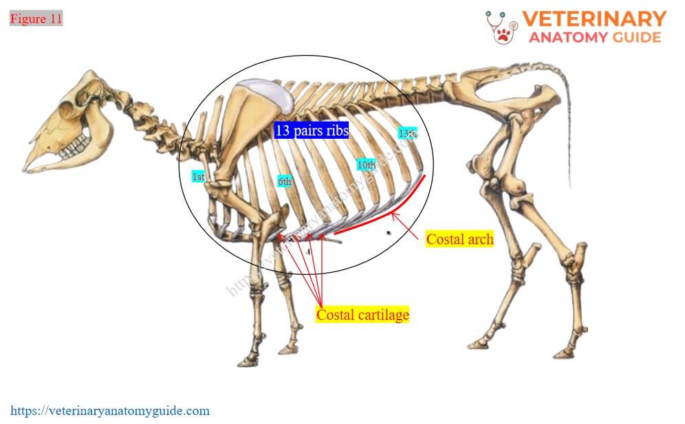
The number of ribs is specific, and you will find 13 pairs of ribs (thirteen) in the cow. Typically, the ribs of animals are of three types –
- Sternal ribs (true ribs): They are connected to the sternum directly through their own costal cartilage.
- Asternal ribs (false ribs): They are connected to the sternum indirectly through their cartilage, which joins with the preceding costal cartilage.
- Floating ribs: these ribs have no connection with the sternum (directly or indirectly).
The first 8 pairs of ribs in the cow are sternal, and the last 5 pairs are asternal. You will not find any floating ribs in the thoracic cage of the cow.
A typical rib of a cow has 2 extremities (proximal and distal) and a body. From the cow rib, you might identify the following osteological features –
- Head of the rib (has 2 articular facets),
- Neck (constricted part between head and tubercle),
- Tubercle of the rib (has 1 articular facet),
- Cranial and caudal border of the body,
- Lateral and medial surfaces of the rib’s body,
- Costal groove on the caudo-medial aspect of the body, and
- The distal end connects the costal cartilage,
What is the costal cartilage and costal arch?
No ribs are directly connected to the sternum; rather, they connect to the sternum with the help of cartilage. These cartilages are known as the costal cartilage.
So the costal cartilages have two articular ends – one for the distal end of the ribs, another one for the facet of the sternum.
All the asternal ribs of the cow’s thorax connect to each other and form a curved structure; this structure is known as the costal arch.
Figure 11 shows the different features of the cow’s rib, along with the costal cartilage and also costal arch.
Cow’s sternum identification
The sternum of a cow is a long plate and osteo-cartilaginous structure. It consists of 7 sternebrae or segments and forms the floor of the cow’s thoracic cavity.
The first segment of the sternum is known as the manubrium, and the last one is the xiphoid cartilage. You will find seven pairs of sternal facets on the cow’s sternum. The first pair of facets of the sternum is located on the manubrium.
For description purposes, the cow sternum consists of two extremities (cranial and caudal) and a body. The body of the sternum possesses two surfaces (dorsal and ventral) and two borders (right and left lateral).
The dorsal surface of the cow’s sternum is wide and covered with the superior ligament and muscle. Again, the ventral surface is little concave and gives origin to the pectoral muscles.
Bones of the forelimb from a cow skeleton
Before going to identifying the bones of the forelimb from the cow skeleton, you might know the segments of the forelimb. Table 3 shows the segments of the cow’s forelimb along with their contents or bones –
| Segments of the cow’s forelimb | Bones |
| Thoracic or shoulder girdle | Scapula, clavicle, and coracoid |
| Brachium or arm region | Humerus bone |
| Antebrachium or forearm region | Radius and ulna bones |
| Manus consists of – Carpus or knee Metacarpus (cannon and splint) Phalanges (digits) | 6 carpal bones Large and small metacarpals Proximal, middle, and distal phalanges, proximal sesamoids, and distal sesamoids |
Figure 12 shows all the bones of the forelimb along with the different segments.

In cows, the clavicle is absent, and the coracoid process is underdeveloped. Now, let’s identify the bones from the cow’s forelimb with their main identifying features. But, you may learn the details anatomical features of the forelimb bones of a cow from the articles mentioned below –
| Scapula bone | Cow scapula bone anatomy |
| Humerus bone | Cow humerus bone anatomy |
| Radius and ulna bones | Cow radius and ulna bones anatomy |
| Carpus | Cow carpus anatomy |
To identify the bones (not including the osteological features) from the forelimb of a cow, you might consider the following –
- First, the type and shape of the specific bone,
- Second, the direction of that bone,
- Third, two or more unique osteological features of the respective bone,
Cow scapula bone identification
The scapula of a cow is a triangular, flat bone of the forelimb. It is also known as the shoulder blade, and the main identifying features are –
- Type and shape of bone: flat and triangular bone,
- Bone direction: downward and forward (ventral and cranial),
- Spine: the lateral surface bears a spine that divides this surface into two portions,
- Acromion process: the distal part of the spine possesses an acromion process.
- Proximal extremity: bears the dorsal border and scapular cartilage,
- Distal extremity: it contains the glenoid cavity, glenoid notch, tuber scapulae, and coracoid process (medially),
Humerus bone of the cow identification
After the scapula bone, the next bone of the cow’s forelimb is the humerus. The key identifying features of the cow’s humerus bone are –
- Type and shape of the bone: long bone (having 2 extremities and a body), and cylindrical shape.
- Direction of the bone: obliquely downward and backward (ventral and caudal),
- Musculospiral groove: it is present on the lateral surface of the body,
- Humeral crest: located between the lateral and cranial surfaces,
- Proximal extremity: the proximal extremity of the cow’s humerus bone contains the head, neck, tuberosities, and bicipital groove.
- Distal extremity: contains radial fossa (cranial) and olecranon fossa (caudally),
Cow’s radius and ulna bones identification
The radius and ulna bones in the cow’s forelimb are fused with each other. The main identifying features of the cow’s radius and ulna bones are –
- Type and shape of the bone: radius (long bone), and ulna (modified, ill-developed long bone),
- Direction of the bone: vertical direction,
- Attachment: the ulna bone is fused caudo-lateral aspect of the radius bone.
- Interosseous space: present between the caudal surface of the radius and the cranial surface of the ulna bone,
- Radial tuberosity: located on the cranio-medial aspect of the proximal part of the radius bone,
- The distal part of the radius present three articular facets for the radial, intermediate, and ulnar carpal.
- Olecranon process: present in the proximal part of the ulna bone (proximally),
- Styloid process:it is the pointed distal part of the ulna bone,
Carpal bones of the cow
There are 6 (six) carpal bones in the cow carpus, or knee, and arranged in two rows. In the first row, you will find radial, intermediate, ulnar, and accessory carpal bones (medial to lateral).
Again, the second row contains the second and third fused bones, and the fourth carpal bone (medial to lateral).
The first row carpal bones make a joint with the distal end of the radius and ulna (where you have already found 3 articular facets for radial, intermediate, and ulnar carpals). The accessory carpal bone is placed laterally and attaches to the articular surface of the ulnar carpal bone.
Again, the distal row carpal bone makes a joint with the proximal end of the metacarpal bone (next bone). As the second and third fused carpals are larger, they will articulate a larger articular surface of the metacarpal bone.
If you notice, the proximal end of the large metacarpal bone present two articular surfaces – larger (medially) and smaller (laterally). You may easily identify the right or left metacarpal by considering this feature (proximal large articular surface faces medially).
Figure 13 shows the arrangement of the cow’s carpal bones. It also presents the proximal and distal articulation with the distal end of the radius, ulna, and large metacarpal, respectively.

Cow’s large and small metacarpal bones
There are two types of metacarpals – large and small metacarpals in a cow. The large metacarpal (also known as the cannon bone) of a cow is a fusion of metacarpal III and IV.
The dorsal or palmar longitudinal median groove indicates the fusion of metacarpal III and IV. It presents a body and two extremities (proximal and distal).
The key identifying features of the large metacarpal of a cow are –
- Type and shape of the bone: long bone and cylindrical (modified),
- Direction of the bone: vertical (downward),
- Proximal end: bears 2 articular surfaces (medially – large, and laterally – smaller) for the distal row of carpal bones,
- Longitudinal median groove: present dorsal (prominent) and ventral longitudinal median groove,
- Distal end: present condyles (lateral and medial), and intercondyloid cleft,
- Facets for proximal sesamoid bones: at the caudal aspect of the distal part of the metacarpal,
The small metacarpal is II and attaches to the caudolateral aspect of the large metacarpal bone in a cow. It possesses a broad proximal end and a narrow distal end.
Phalanges of the cow’s forelimb
The number of developed digits in any animal corresponds to the number of developed metacarpals. As the cow has two developed metacarpal (III & IV), there are 2 developed digits in each forelimb.
The developed digits of a cow are – III and IV, and each consists of three phalanges –
- Proximal phalanx (first phalanx): It is elongated and directed downward and forward. The body of the proximal phalanx possesses 4 surfaces, where the dorsal and lateral surfaces are continue.
- Middle or second phalanx: it is shorter and directed downward and forward. The body of this phalanx possesses three surfaces, where the lateral surface is rounded and irregularly concave.
- Distal phalanx (third phalanx): It is roughly triangular in shape and possesses 4 surfaces.
Between the middle and distal phalanx, there is one sesamoid bone in the cow. This is the distal sesamoid bone (also known as the navicular bone) in the cow’s forelimb.
In cows, the second and fifth digits are rudimentary and known as the dew-claw. They are located at the palmar aspect of the metacarpo-phalangeal joint without forming any joint with any bones.
Proximal and distal sesamoid bones in the cow’s forelimb
You will find 4 oval-elongated sesamoid bones in each forelimb of a cow at the proximal metacarpophalangeal joint on its palmar aspect. There are two for each developed digit, and the axial one is larger than the lateral.
Again, there are two sesamoid bones in the cow’s forelimb, one for each developed digit. They are almost flat, small bones located transverse between the middle and distal phalanges.
Figure 14 shows the three phalanges, the proximal sesamoid, and the distal sesamoid bones.

Bones of the hindlimb from a cow skeleton
The bones of the hindlimb from a cow skeleton include – hip bone, femur, tibia, fibula, tarsals, metatarsals, sesamoids, and phalanges. But, before going to identify these bones from the cow’s hindlimb, you might know its segments.
Table 4 shows the segments of the cow’s hindlimb with their contents or bones –
| Segments of the cow’s hindlimb | Bones |
| Pelvic girdle | Sacrum, pelvis: ilium, ischium, pubis |
| Thigh region | Femur bone |
| Crus or true leg region | Tibia and fibula bones |
| Pes region includes – Tarsus or hock region Metatarsus (cannon and splint)Digits (phalanges) | 5 tarsal bonesLarge and small metatarsals 3 phalanges, proximal sesamoids, and distal sesamoids |
Figure 15 shows the segments of the cow’s hindlimb along with their bones.

All the bones from the cow’s hind limb possess huge, unique osteological features. You may learn all the bones individually with lots of resources from the articles mentioned below –
| Hip bone or os coxae | Cow hip/ cow pelvic bone anatomy |
| Femur bone | Cow femur bone anatomy |
| Tibia and fibula | Cow tibia and fibula bones |
| Tarsal, metatarsal, and phalanges | Cow pes anatomy |
To identify all the bones (not including the osteological features), you might consider the following –
- Type and shape of the hindlimb bone,
- Direction of the hindlimb bone, and
- Two or more identifying osteological features,
Cow hip bone (os coxae)
There are right and left hip bones in a cow that join each other through the pelvic symphysis. Each of the hip bones (also known as the os coxae) of a cow consists of the ilium, ischium, and pubis.
It is very hard to identify the practical extension of the ilium, ischium, and pubis. But the larger acetabulum can be considered as the landmark to identify roughly the ilium, ischium, and pubis.
The ilium, ischium, and pubis fuse to form the larger acetabular cavity in a cow, where the head of the femur attaches. The cranial triangular flat part of the hip is termed the ilium bone. Again, the caudal roughly quadrilateral plate is known as the ischium bone. Finally, the ventral and craniomedial small triangular part of the hip is the pubis bone.
Figure 16 shows the ilium, ischium, and pubis (both right and left) of the cow’s hip bone.

A few key features of these cows’ hip bones are –
- Type and shape of the bones: all are flat bones; the ilium is triangular, the ischium is quadrilateral, and the pubis is small and triangular.
- Ilium bone: possesses the tuber coxae, the tuber sacrale, the gluteal line, and the greater ischiatic notch.
- Ischium bone: it contains the greater ischiatic spine, the lesser ischiatic notch, the ischial tuberosity, and forms the ischial arch caudally.
- Pubis bone: possesses the ilio-pubic eminence and forms the pelvic floor with the ischium bones.
The pelvic symphysis consists of the pubic symphysis (between the right and left pubic bones) and the ischial symphysis (right and left ischium bones). Again, the oval elongated obturator foramen is formed between the pubic and ischium bones of the cow’s hip.
Cow femur bone from the hindlimb
The femur is the largest bone in a cow’s skeleton. Proximally, it contains a head that is attached to the acetabular cavity (cotyloid cavity).
The key features for identifying the cow femur bone (also known as the round bone) from its hindlimb are –
- Type and shape of the bone: the largest long bone, with a cylindrical shape.
- Direction of the bone: downward and forward (oblique),
- Proximal extremity: contains head with fovea capitis femoris (medially), greater trochanter (laterally), trochanteric ridge (caudo-laterally), and trochanteric fossa (caudally),
- Distal extremity: present trochlea (cranially), and lateral and medial condyles (caudally),
The trochanteric ridge in the cow’s femur bone is oblique (extends from the lateral to the medial aspect). In front of the trochlea of the femur, you will find a triangular sesamoid bone (patella).
Figure 17 shows the proximal and distal extremities of the cow’s femur with their osteological features. It also shows the location of the patella (proximal triangular sesamoid) on the groove of the trochlea of the cow’s femur bone.

Tibia and fibular bones identification from a cow’s hind limb
The tibia of a cow is a strong and massive bone in its hindlimb. Again, the fibula is a highly rudimentary bone in the cow, and its proximal end attaches to the lateral condyle of the tibia.
The main identifying features of the cow tibia are –
- Type and shape of the bone: long bone (prismatic at upper end, narrow and flat at distal end),
- Direction of the bone: downward and backward directed,
- Lateral surface of tibia: concave and gradually spirals down to the front,
- Tibial crest: it is located at the proximal part of the cranial border (very prominent),
- Distal extremity: contains two grooves for articulation with the ridge of the tibial tarsal (talus),
- Popliteal line: present at the caudal surface of the tibia (extends from the lateral to the medial surface),
Cow tarsal bones identification
There are 5 tarsal bones in the tarsus of a cow’s hindlimb. They are the short bones located between the distal extremity of the tibia bone and the proximal extremity of the metatarsal bone.
These cows’ tarsal bones are arranged into 3 rows – proximal, middle, and distal –
- Proximal row: this row contains the tibial tarsal (talus) and fibular tarsal (calcaneus). The calcaneus is located lateral to the tibial tarsal or talus.
- The middle row: it contains the fused central tarsal and fourth tarsal. This fused tarsal joins with the lateral articular surface of the metatarsal (lateral aspect).
- Distal row: this row contains the small first tarsal and fused second and third tarsals (they are arranged from medial to lateral). The fused second and third tarsals articulate with the medial articular surface of the metatarsal bone (medial aspect).
The tibial tarsal (talus) is in the form of an elongated pully and possesses 2 ridges at the proximal end and 2 trochlea at the distal end. Again, the fibular tarsal (calcaneous) is attached to the caudolateral aspect of the tibial tarsal (talus).
The proximal ridges of the tibial tarsal are vertical in the cow. However, the fibular tarsal (calcaneus) is an elongated short bone placed obliquely.
Metatarsal bones in a cow
Cows also possess the large and small metatarsal bones. But the large metatarsal is the fusion of metatarsal III and metatarsal IV. Again, the cow’s small metatarsal is II that attaches to the medial aspect of the large metatarsal.
Like the metacarpal, the cow’s metatarsal also possesses two extremities (proximal and distal), and a 4-sided body. Again, you will find almost similar osteological features in the cow’s large metatarsal, like metacarpals.
There are some differentiating osteological features in the cow’s large metatarsal compared to the large metacarpal –
- Length: the length of the cow’s large metatarsal is longer compared to the metacarpal.
- Surfaces in the body: there are 4 surfaces (lateral, medial, dorsal, and palmar) in the cow’s large metatarsal, whereas the metacarpal possesses only 2 surfaces (dorsal and palmar),
- Dorsal longitudinal groove: It is deeper in the cow’s metatarsal bone compared to the metacarpal.
- Proximal medial articular surface: it is smaller in the cow’s metatarsal.
The small metatarsal of a cow is rudimentary and quadrilateral in form. This bone articulates with the cranio-medial aspect of the large metatarsal (proximal).
Figure 18 shows the tarsals, metatarsal bones from the cow’s hindlimb. It also presents the phalanges and sesamoid bones of the cow’s hindlimb.

The phalanges and sesamoid bones in the hindlimb are almost similar to the cow’s forelimb.
Visceral bones in a cow
There are two small triangular bones found in the myocardium of the cow’s heart. But these visceral bones are not part of the cow skeleton. They are located on either side of the aortic ring of the cow’s heart.
Cow skeleton images and diagrams
In this part, I will provide some of the images and labeled diagrams of the cow skeleton. These images and labeled diagrams represent the bones from different regions of the head, vertebrae, thorax, and limbs.
Figure 19 shows the different bones from the cow skeleton.
Conclusion
So, the cow skeleton provides the strong framework to its body and consists of bones, cartilage, and ligaments. The beginner students might identify these bones from the axial and appendicular skeleton perfectly.
This descriptive guide might help the beginner identify all of these bones from different regions of the cow’s skeleton. They can now learn the detail osteological features of every single bone in the skeleton.
References
- Mcleod, W. N.: Bovine anatomy, 2nd Edn. Burgess publishing co. Minneapolis.
- Ghosh, R. K. : Primary veterinary anatomy, 2nd Edn.Current Books International, Kolkata.
- Rowen D. Frandson, W. Lee Wilke, and A. Dee: Anatomy and physiology of farm animals, 4th edn.Wiley-Blackwell, USA.
- Hermanson et al., Miller and Evan’s Anatomy of the dog, 5th Edn. Elsevier.
- Getty, R,. Sission and Grossman’s The anatomy of the domestic animals, 5th Edn. W. B. Saynders and Co. Philadelphia (USA).
- Nickel, R. Schummer, A. and Seiferle, E.: The anatomy of domestic animals, 1st Edn. Verlag Paul Parey, Berlin.
- Dyce, K. M., W. O. Sack and CSG Wensing : Text Book of Veterinary Anatomy, Lea & Febiger. Philadelphia
