

The cow humerus is the first long bone that forms the arm segment of the forelimb. In this guide, I will help you to learn the osteological features of the cow humerus bone with different learning resources.
Quick answer: The cow humerus possesses a cylindrical shaft and two expanded extremities: proximal and distal. Again, the shaft is twisted and presents four distinguished surfaces with different unique features.
You will learn the osteological features from these four surfaces and also from the two extremities. Finally, I will also help you to identify the right / left humerus bones from the cow’s forelimbs.
Cow humerus
The cow humerus is classified as a long type of bone. It articulates with the scapula above and the radius and ulna bones below.
This is the first time that I am going to describe osteological features from the long bone of a cow skeleton. Thus, first, you might know what you should learn from every long bone and how to describe it perfectly.
While studying long bones like the humerus, you might know the followings –
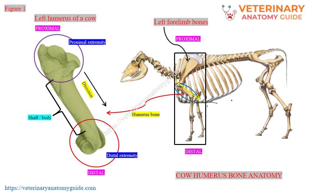
- Location of the bone: the humerus is located between the scapula and the radius and ulna bones. It forms the arm segment of the forelimb of a cow.
- Direction of the bone: This bone is directed obliquely downward and backward.
- Shaft and extremities: Every long bone, like the humerus, possesses a cylindrical shaft / body and two extremities (proximal and distal).
- Surfaces of the shaft: the shaft of the humerus bone consists of four surfaces (anterior, posterior, lateral, and medial). You might know the unique identifying features of these surfaces.
- Extremities of the bone: the long bone, like the humerus, consists of a proximal and distal extremity. Here, the unique features of the proximal extremity of the humerus are the presence of the head, neck, and tuberosities. Again, the distal extremity possesses condyles, epicondyles, radial fossa (cranial), and olecranon fossa (caudal).
Here, Figure 1 shows the location, direction, shaft, and extremities of the cow’s humerus bone from its forelimb.
[For each long bone of the cow skeleton, you might follow these learning strategies with little or no changes. The osteological features of the four different surfaces and extremities are unique in each long bone.]
Cow humerus bone identification
Here, I would like to list the osteological features that you will find 4 different surfaces of the shaft and two extremities. This might help you to describe the features of the humerus bone perfectly.
Osteological features of the shaft of the humerus
- Lateral / external surface: Spiral and present a musculospiral groove.
- Medial / internal surface: rounded and presents a teres tubercle at the middle.
- Anterior / cranial surface: triangular, wide and smooth above, and narrow below.
- Posterior / caudal surface: smooth and rounded.
- Humeral crest: forms between the anterior and lateral surface (cranially).
- Deltoid tuberosity: at the middle of the humeral crest.
- Proximal extremity: present a expanded head, constricted neck, and two tuberosities (lateral and medial).
- Bicipital / intertubercular groove: forms between the lateral and medial tuberosity (cranially).
- Distal extremity: present lateral and medial condyles and epicondyles.
- Coronoid / radial fossa: It is located cranially and above the condyles.
- Olecranon fossa: It is located caudally and is a deep fossa.
Here, Figure 2 shows all the above -mentioned osteological features from the shaft and extremities of the cow’s humerus bone.

[For all bones / long bones, first you should identify their key identifying features from different surfaces and extremities. Then you should learn the details or description of these osteological features one by one.]
Cow humerus bone anatomy
Now, I will describe all the osteological features of the shaft and extremities of the cow’s humerus bone in detail. First, let’s start with the features from different surfaces of the shaft / body of the humerus bone.
The shaft of the cow’s humerus bone
The shaft / body of the cow’s humerus bone is irregularly cylindrical shape. It has a twisted appearance and presents four (4) different surfaces –
- External / lateral surface,
- Internal / medial surface,
- Anterior / cranial surface, and
- Posterior / caudal surface,
External surface of the cow’s humerus
This external or lateral surface of the cow’s humerus bone is smooth and spiral. Thus, this surface forms a musculospiral groove that contains the brachialis muscle.
The musculospiral groove is the key identifying feature of the lateral surface of the shaft of the cow’s humerus bone. With this feature, you may easily identify the right / left humerus bones from the cow’s forelimb.
Here, Figure 3 shows the musculospiral groove from the lateral surface of the shaft of the cow humerus bone.

This external surface of the humerus bone continues with the posterior surface above. Again, it continues with the cranial border (humeral crest) below.
Internal / medial surface of the cow’s humerus
The internal or medial surface of the shaft of the cow’s humerus is nearly straight and rounded from side to side. This surface blends with the posterior and anterior surfaces of the shaft.
In the middle of the internal border, there is a teres tubercle. Within this tubercle, the tendons of the teres major and lattissimus dorsi muscles insert.
Here, Figure 4 shows the teres major tubercle on the medial / internal surface of the shaft of the cow’s humerus bone.

You will also find the nutrient foramen at the lower third part of this internal surface of the humeral shaft.
Anterior surface of the cow’s humerus
The anterior surface of the shaft of the cow’s humerus bone shows the followings –
- Upper segment: the upper segment of the anterior surface is a triangle, smooth, and wide.
- Lower segment: This lower segment is narrow and rough.
The anterior surface of the humeral shaft is separated from the external surface by a distinct ridge / border. This ridge or border is known as the humeral crest of the cow’s humerus bone.
In the middle of the humeral crest on its upper part, there is a rough prominence. This rough prominence is the deltoid tuberosity of the humerus bone.
The deltoid muscle (DM) inserts on the deltoid tuberosity of the humerus. Here, Figure 5 shows the deltoid tuberosity and the anterior surface of the cow’s humerus bone.

From the deltoid tuberosity, a rough line curves upward and backward to the external surface of the neck (shown in the figure). This curve line gives the origin of the lateral head of the triceps brachii (3 heads) muscle in cows.
Again, the lower part of the crest of the humerus becomes less prominent. Finally, it ends on the coronoid fossa at the cranial distal part of the distal extremity of the humerus bone.
Posterior surface of the humerus shaft
The posterior / caudal surface of the shaft of the cow’s humerus bone is rounded from side to side. This surface is relatively smooth and possesses nutrient foramina at the distal third portion.
Extremities of the humerus bone of a cow
You will find different key identifying osteological features on the proximal and distal extremities of the cow humerus bone. These features also help you to identify the right / left bones as well as to identify the proximal and distal parts.
Proximal extremity of the cow’s humerus
The proximal extremity of the cow’s humerus bone contains an expanded head, a narrow neck, two tuberosities, and a bicipital groove. Here, the head possess an almost circular convex articular surface.
It is very extensive and articulates with the glenoid cavity of the cow’s scapula bone above. There is a fossa (depression) in front of the head that contains several foramina.
The neck is the constricted part below the head. In humerus, the neck is located behind and below the head.
Figure 6 shows the osteological features from the proximal extremity of the cow’s humerus bone.

Tuberosities from the proximal end of the humerus bone
There are two tuberosities in the proximal end of the cow’s humerus bone: external and internal. Here, the external or lateral tuberosity is located on the anterior / cranial and lateral aspect of the proximal extremity.
This external / lateral tuberosity is comparatively larger and more prominent. It is divided into two parts – cranial and caudal.
The external tuberosity forms the lateral boundary of the bicipital groove. This area gives attachment to the supraspinatus and infraspinatus muscles. Again, the bicipital groove gives passage to the tendon of the biceps muscle.
The internal / medial tuberosity is less developed and also consists of two parts – cranial and caudal. Here, the cranial / anterior part of the internal tuberosity forms the inner boundary of the bicipital groove.
The bicipital groove is primarily covered by the cartilage in a fresh state. Finally, it attaches to the tendon of origin of the biceps brachii muscle.
Distal extremity of the cow humerus bone
The distal extremity (lower part) of the cow humerus bone possesses the articular surfaces for articulation with the radius and ulna bones. This extremity consists of two unequal and modified condyles – internal and external.
It also consists of internal and external epicondyles, the posterior olecranon fossa, and the cranial radial / coronoid fossa. Here, the internal condyles are known as trochlea, and the external condyle is known as capitulum.
Figure 7 shows the different osteological features of the distal extremity of the cow’s humerus bone.

Condyles of the humerus
The internal condyle (trochlea) is larger and traversed by a ridge. This groove extends cranially to the radial fossa and also caudally to the olecranon fossa.
Here, the radial fossa / coronoid fossa is located in front of the groove of the internal condyle. It furnishes the origin of the extensor carpi and common extensor muscles of the forearm.
Between the condyles at the posterior aspect, the deep cavity is the olecranon fossa.
The radial fossa accommodates the coronoid / condyloid process (sagittal ridge) of the radius bone during flexion. Again, the olecranon process accommodates the anconeus process of the ulna bone during extension of the cow’s forelimb.
Epicondyles of the humerus bone
You will find the internal and external epicondyles from the corresponding condyles at the distal extremity. They are the small projections from the corresponding condyles of the humerus bone.
The internal epicondyle is small and furnishes origin to the flexor muscles of the carpus and digits. Again, the external epicondyle gives origin to the extensor carpi muscle.
You will find a supracondyloid crest above the external epicondyle of the humerus bone. From this supracondyloid crest, the lateral boundary of the musculospiral groove is formed.
Figure 8 shows the external and internal epicondyles and supracondyloid crest from the distal extremity of the cow’s humerus bone.

How to differentiate the cow humerus from the horse, dog, and pig humerus?
Well, this is very important to differentiate the cow’s humerus bones from those of horses, dogs, and pigs at your anatomy learning laboratory. You may easily differentiate the cow’s humerus bone from other species by following the below-mentioned information.
Horse humerus bone unique features
The shaft of the horse’s humerus bone is more twisted than that of the cow’s. It present the more deep musculosprial groove on its lateral surface compared to the cows.
Again, the deltoid tuberosity of the horse’s humerus bone is more prominent than that of the cow. The curve line above the deltoid tuberosity is more distinct in the horse humerus.
You will find three tuberosities at the proximal end of the horse’s humerus, where the cow’s humerus shows two tuberosities. In the horse humerus, there are external, intermediate, and internal tuberosities (tubercles). Thus, the bicipital groove is divided into two parts.
Again, the radial and olecranon fossa are more deep in the horse humerus bone compared to the cow’s. Here, Figure 9 shows the comparative osteological features of the humerus bones between the cows and horses.

Dog humerus bone unique features
The shaft of the dog humerus bone is less twisted and long that of the cows. Here, the musculospiral groove is also less prominent in the dog’s humerus.
The deltoid tuberosity is in the ridge form in a dog’s humerus. Again, the curved line above the deltoid tuberosity is very indistinct.
The head of the dog’s humerus bone is more rounded and convex than that of the cow’s. It shows external and internal tuberosities on the proximal extremity, like the cows.
The most unique feature found in the distal extremity of the dog humerus bone is You will find a supratrochlear foramen that communicates between the radial and olecranon fossa.
Here, Figure 10 shows the comparative anatomy of the humerus bones between the dogs and cows.

Pig humerus bone anatomy: unique features
The shaft of the pig humerus bone is a little different from that of the cow’s humerus. It is less twisted and possesses a shallow musculospiral groove.
Both the proximal and distal extremities of the pig humerus are slightly inclined. You will find the supratrochlear foramen in the pig humerus bone, which also communicates with the radial and olecranon fossa.
Here, Figure 11 shows the features of the pig humerus bone compared to those of the cow.

How to identify the right / left humerus bone in a cow?
Well, you might identify the right / left humerus bone in a cow while studying their features at laboratory. To identify the right / left humerus from the cows, you might have basic knowledge of their osteological features.
If you want to identify the right / left humerus from the cow’s forelimb bones, you might consider the followings –
- First, identify the proximal and distal extremities. Here, the proximal extremity contains the head, neck, and tuberosities, while the distal extremity contains the condyles, epicondyles, radial, and olecranon fossa.
- Second, hold the bone in an oblique direction first so that the head faces proximally. The head will remain caudally and medially, where the larger tuberosity will faces lateral.
- Third, make sure that the musculospiral groove from the external surface of the shaft faces laterally.
- Fourth, also make sure that the radial fossa faces cranial, whereas the olecranon fossa faces caudal.
- Fifth, now hold the bone with the right / left hand according to the direction of the bone as it is found in the forelimb in a normal standing condition.
Now, it’s time to verify the followings –
- Head: faces proximally and caudomedially.
- Condyles: faces distally and cranially,
- Radial fossa: faces cranially,
- Olecranon fossa: faces caudally,
- Musculospiral groove: faces lateral to the shaft,
- Humeral crest: faces cranially / anteriorly,
- Direction: oblique (downward and backward).
If this bone meets the above-mentioned features while holding it with the right and left hand, then it will be the right / left bone, respectively.
Here, Figure 12 shows the identification technique for identifying the right / left humerus bone in cows.

Why is this humerus bone the right one?
Figure 13 shows the right humerus bone of a cow. Let’s explain why this is the right humerus bone of the cow’s.

First, let’s hold this bone with the left hand and make sure the head faces proximal, and the condyle faces distal. Now, you will observe the followings –
- Head: faces proximal aspect,
- Condyle: faces the distal aspect,
- Musculospiral groove: faces medial / internal aspect,
But the musculospiral groove is present on the lateral / external surface of the shaft. Thus, this is not the left side humerus bone of the cow’s forelimb.
Now, let’s hold the bone with the right hand while the head and condyles face the proximal and distal aspect respectively. You will observe the followings from the bone –
- Head and condyles: face the proximal and distal aspects, respectively.
- Musculospiral groove: faces the lateral / external surface of the shaft.
- Crest of humerus: faces the anterior aspect.
Thus, this humerus bone is the right side bone of the cow’s forelimb.
[So, if you want to identify the right / left bones from the animal’s limb, you might consider some features as landmarks. Again, the direction of the bone in normal standing conditions is essential to hold the bone perfectly with the right / left hand.]
Conclusion
So, the cow humerus bone possesses a twisted and cylindrical shaft and two expanded extremities. Here, the twisted and cylindrical shaft possesses four different surfaces with unique osteological features.
The lateral musculospiral groove and deltoid tuberosity are the key identifying features of the humeral shaft. Again, the anterior crest of the humerus is another identifying feature of the cow’s humerus bone.
The proximal extremity shows the expanded head, narrow caudal neck, and two disticnt tubersity with bicipital groove. Again, two modified condyles, radial fossa, and olecranon fossa are unique osteological features of the distal extremity of the cow’s humerus bone.
References
- Singh, G et al., Comparative morphological and morphometrical analysis of the humerus bone in bovines: A study of Ox (Bos indicus) and Nilgai (Boselaphus tragocamelus). Inter. Jr. Vet. Sci. Ani Hus. 2024; 9(2): 941-946
- Ghosh, R. K. : Primary veterinary anatomy, 2nd Edn.Current Books International, Kolkata.
- Rowen D. Frandson, W. Lee Wilke, and A. Dee: Anatomy and physiology of farm animals, 4th edn.Wiley-Blackwell, USA.
- Raghavan D. Anatomy of Ox. Indian Council of Agricultural Research (ICAR), ND, India; c1964.
- Best Adam et al., Anatomy, histology, development and functions of ossa cordis, Anatomia, histologia, embryologia, 2022.
- Getty, R,. Sission and Grossman’s (Getty) The anatomy of the domestic animals, 5th Edn. W. B. Saynders and Co. Philadelphia (USA).
- Choudhary, O et al., Gross and Biometrical Studies on Humerus and Radius Ulna of Chital (Axis axis). Indian Journal of Veterinary Anatomy,2013; 25(1).
- Nickel, R. Schummer, A. and Seiferle, E.: The anatomy of domestic animals, 1st Edn. Verlag Paul Parey, Berlin.
- Larsen et al., 2023. Navicular bone fracture and severe deep digital flexor tendinopathy after palmar digital neurectomy in two horses. Open Veterinary Journal, 13 (12).
- Talukdar M, et al., Gross Anatomical study on the humerus of Mithun. Indian Veterinary Journal. 2002;79:585-587.
- Hermanson et al., Miller and Evan’s Anatomy of the dog, 5th Edn. Elsevier.
- Mcleod, W. N.: Bovine anatomy, 2nd Edn. Burgess publishing co. Minneapolis.
- Sarma K, Kalita SN, Kumar P. Anatomy of the humerus of Nilgai (Boselaphus tragocamalus). Indian Journal of Animal Sciences. 2008;78(1):24-27.
- Dyce, K. M., W. O. Sack and CSG Wensing : Text Book of Veterinary Anatomy, Lea & Febiger. Philadelphia
