
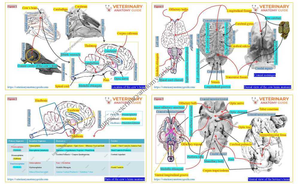
Cow brain anatomy refers to the features of its three major parts. It is part of the cow’s central nervous system and is considered the most complex organ.
In this guide, I will describe the anatomical features of the cow’s brain with hand-drawn diagrams and real pictures.
Quick answer: The cow brain is the body’s command center, and its anatomy includes the forebrain, midbrain, and hindbrain.
I will help you understand every feature of the cow’s brain with an easy learning technique. This guide on the cow’s brain will help you prepare for both theory and practical examinations with confidence.
Where is the cow’s brain located?
Figure 1 shows the location of the cow’s brain within the cranial cavity. This hand-drawn figure provides an overview of different parts of the brain and their relationships with surrounding organs.
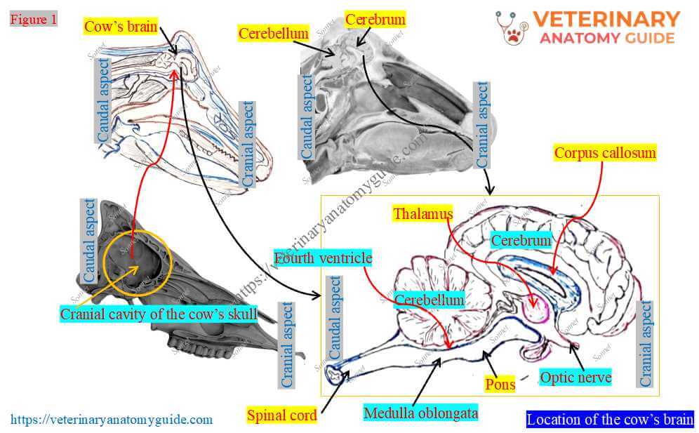
Location: The cow’s brain is located in the oval, elongated, irregular cranial cavity formed by the skull bones. The meninges cover it and continue caudally with the cow’s spinal cord.
In recent years, I have found that students consider it one of the hardest topics in the nervous system. However, I have always tried to present the cow’s brain anatomy efficiently in my practical classes.
Similar strategies are also provided in this guide, so I request that you follow them carefully. Now, I would like to introduce the basic parts (components) of the cow’s brain.
Parts of the cow’s brain anatomy
Figure 2 shows three basic parts (components) of the cow’s brain. But I don’t want you to memorize this first; this is only to familiarize you with the brain’s main parts and subparts.
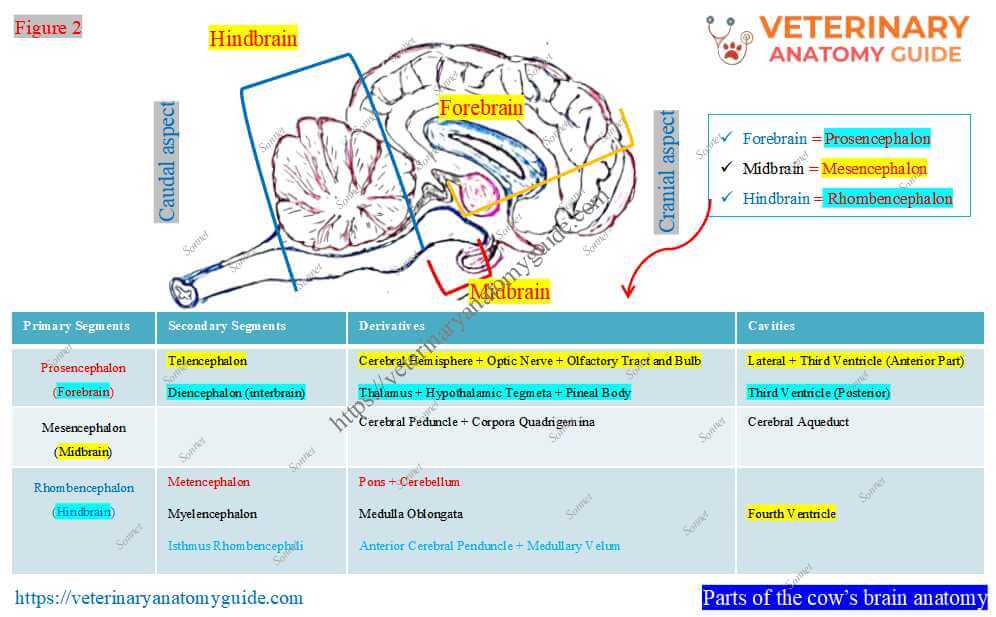
Just try to understand these basic parts well with the help of the above figure. Thus, according to the figure, the following are the major parts of the cow/bovine/cattle brain –
- Forebrain (prosencephalon): This is the rostral part of the brain that consists of the cerebrum, optic nerve, olfactory bulb and tract, thalamus, hypothalamus, and pineal body. However, the cerebrum, optic nerve, and olfactory elements are termed the telencephalon. Whereas, the thalamus, hypothalamus, and pineal body form the diencephalon.
- Midbrain (mesencephalon): It is the middle part of the brain and consists of the cerebral peduncles and corpora quadrigemina. However, the cerebral peduncle and corpora quadrigemina form the cranial part of the brainstem.
- Hindbrain (rhombencephalon): It is the caudal-most part of the brain and comprises the cerebellum, pons, and medulla oblongata. Here, the cerebellum and pons form the metencephalon, whereas the medulla oblongata forms the myencephalon (the caudal part of the brainstem).
Again, these three parts of the cow’s brain contain different ventricles of brain (cavities). A separate description is needed to understand these brain cavities better.
Now, can you tell what you should learn first to complete the cow’s brain anatomy? Once you identify the key features, you can easily classify and describe the parts of the cow’s brain anatomy.
Identification of features from the brain of a cow/bovine/cattle
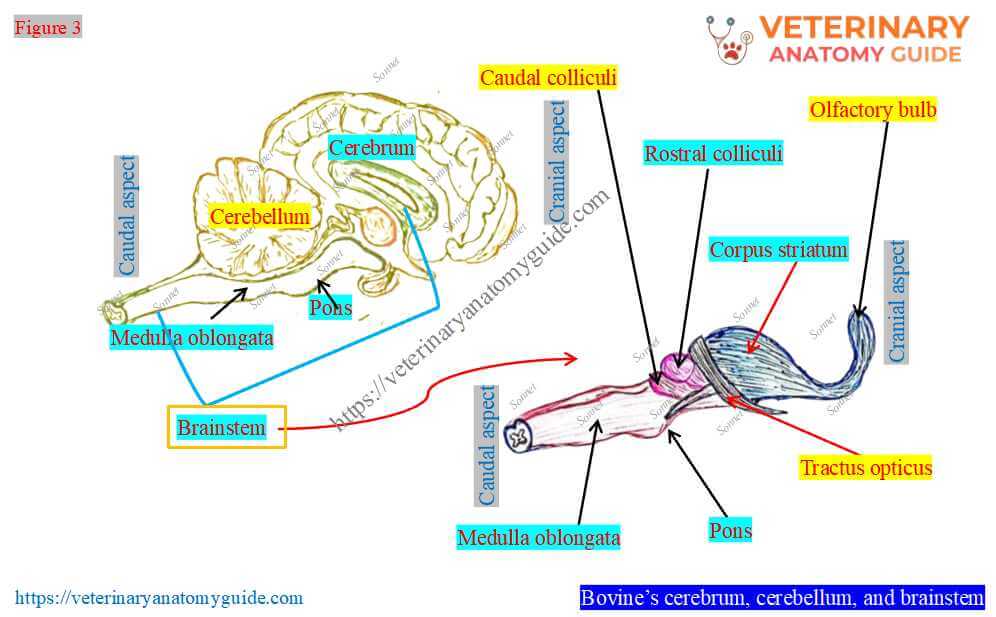
To identify features, I would like to subdivide the cow’s brain into the cerebrum, cerebellum, and brainstem (Figure 3). I will identify features from the dorsal and ventral views of the cow’s whole brain, as well as from the lateral longitudinal view.
Dorsal view of the cow’s brain
Figure 4 presents the anatomical features of the dorsal view of the cow’s brain. I also tried to show the lobes of the bovine cerebrum in this figure.
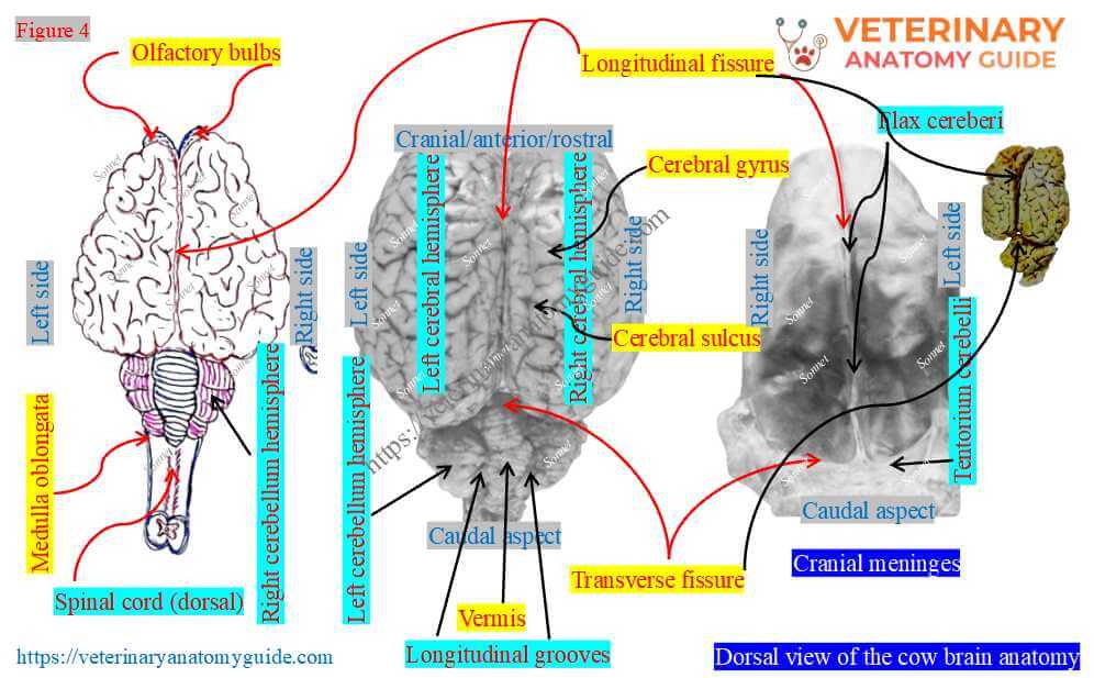
According to the figure, the cranial and largest part of the cow (bovine/cattle) brain is the cerebrum. A longitudinal fissure divides it into two parts: the right and left cerebral hemispheres.
Both cerebral hemispheres have elevated and depressed portions. The elevated part is called a gyrus, whereas the depressed area is called a sulcus.
The figure also highlights that the cerebrum is separated from the caudal part by a transverse fissure. Here, the transverse fissure is the transverse cerebral groove, and the caudal part is termed the cerebellum.
The dorsal surface of the cow’s cerebellum has two longitudinal grooves. These grooves divide the dorsal surface into three segments: the right and left cerebellar hemispheres and the vermis (middle).
However, the dorsal view of the bovine brain also shows the medulla oblongata with a groove. It lies just caudal to the cerebellar hemispheres and continues caudally with the spinal cord.
Ventral view of the bovine’s brain
Figure 5 shows features of the ventral view of the bovine brain. However, similar anatomical features are found on the ventral surface of the brain of sheep, goats, horses, and pigs.
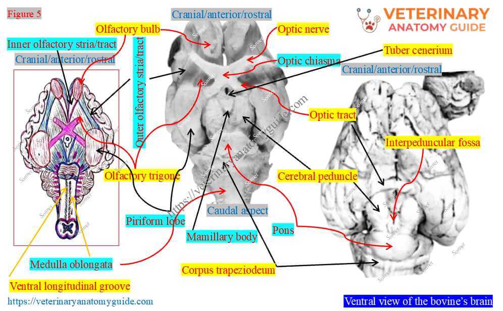
The figure shows elongated structures in the cranial part. These are the olfactory bulbs, which contain the tract or striae caudally.
Just caudal to these bulbs, the figure shows rough triangular areas on both sides (right and left). These are the trigonium olfatorium of the cow’s brain.
Again, caudal to this structure, the ventral surface of the cow’s brain presents elongated raised structures. They are also on both sides and are termed the pyriform lobes.
Medial to each pyriform lobe, the figure highlights another raised area. This is a major brain structure called the cerebral peduncle.
The optic tracts are attached to the corresponding cranial part of the cerebral peduncle. However, the right and left optic tracts cross to form the optic chiasm and then run as individual nerves.
Between the cerebral peduncles is a small mammillary body. Again, cranial to this body, the figure identifies the tuber cinereum.
Caudal to the cerebral peduncles, the figure shows the small elevated area, the pons. It also shows a small area behind the pons, termed the corpus trapezoideum.
A pyramid lies caudal to the trapezoid, but is hard to see in the real sample. Finally, you will find the ventral median groove of the medulla oblongata, which continues into the spinal cord.
Longitudinal view of the cattle (cow) brain
Figure 6 identifies the features from the longitudinal section of the cattle (cow) brain. It also shows some features you have already seen in the dorsal and ventral views.
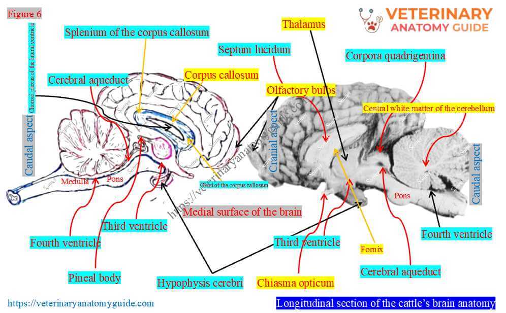
Thus, I tried to highlight only the new features from this longitudinal section of the brain.
The cerebrum part of the cow’s brain presents an elongated hollow structure in the middle. This is the corpus callosum, which presents three parts: genu, splenium, and fornix (identified in the figure).
Here, the rostro-ventral segment of the callosum is the genu. Again, the caudo-dorsal segment is the splenium of the corpus callosum. Finally, the caudal part is the fornix of the corpus callosum.
The figure also highlights the oval structure just caudal to the corpus callosum. It is the cow’s thalamus, which is oval.
Dorsal to the thalamus, the figure identifies a small gland called the pineal gland. However, ventral to the thalamus, the figure also shows the pituitary gland (hypophysis).
Caudal to the pineal gland are two pairs of colliculi: rostral and caudal. You may also view these structures (colliculus and pineal gland) from the dorsal surface of the brain.
In that case, you need to remove the cerebrum or cerebellum hemisphere for best visibility.
Now, caudal to the colliculus, the figure identifies the tree-like structures in the cerebellar section. It also shows a transverse section of the medulla oblongata and spinal cord.
Cavities of the cow’s brain (ventricles)
Figure 7 identifies the cavities (ventricles) and ducts of the cow’s brain.
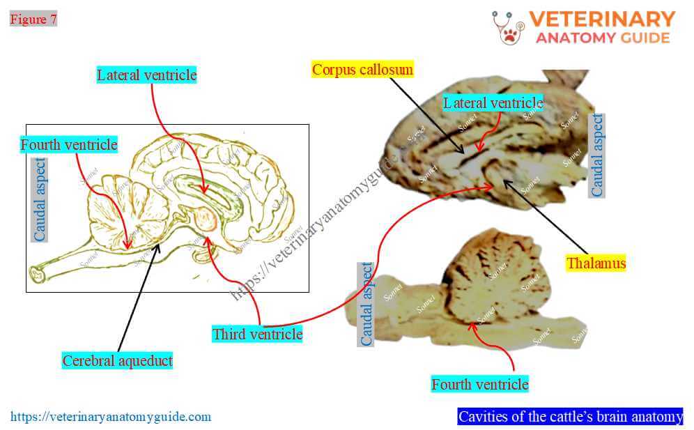
The hollow and elongated corpus callosum forms the lateral cavities. Thus, the cow’s brain has two lateral ventricles.
A septum (called the septum pellucidum) separates the two lateral ventricles (shown in figures 6 and 7).
A space or cavity surrounds the thalamus; this is the third ventricle. However, the space or cavity between the cerebellum, medulla oblongata, and pons is the fourth ventricle.
The figure also identifies the connected duct between the third and fourth ventricles of the cow’s brain. This is the cerebral aqueduct, which is located above the cerebral peduncle.
What is the brainstem in a cow’s brain anatomy?
Figure 8 highlights the brainstem of the cow’s brain anatomy. You may refer to the cow’s brainstem in different ways.
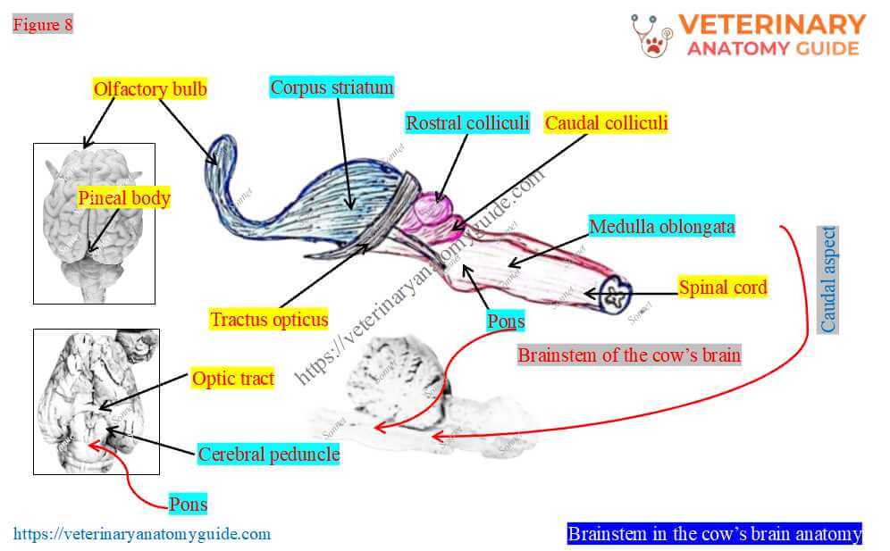
Brainstem: The cow’s brainstem includes the thalamus, hypothalamus, pineal body, cerebral peduncle, colliculus, pons, and medulla oblongata.
Now, you know all of the anatomical features of the cow’s brain. Now, go back to Figure 2, where I show the parts of the cow’s brain.
This figure will help you memorize the divisions and retain your knowledge. Now you are ready to describe the cow brain anatomy completely.
Cow brain anatomy
Figure 9 highlights an overview of the cow’s brain anatomy. From this figure, you can easily see which topics to cover under the anatomy of a cow’s brain.
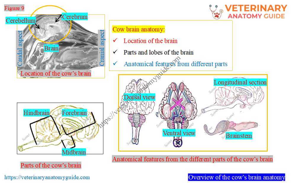
The following are the topics for the complete description of the cow’s brain –
- Location of the cow’s brain
- Parts and lobes of the cow’s brain
- Features, identification, and description of each part of the brain
However, you may describe its covering (cranial meninges) and the ventricular system. It depends on the questions on the cow’s brain in your examination.
Most topics are completed except the description of features from the three major parts. Thus, I will now focus on describing these three parts of the cow’s brain.
Forebrain anatomy (prosencephalon)
Figure 10 highlights only the part of the forebrain (prosencephalon) of the cow’s brain. I will describe the major components of the forebrain with the diagram.
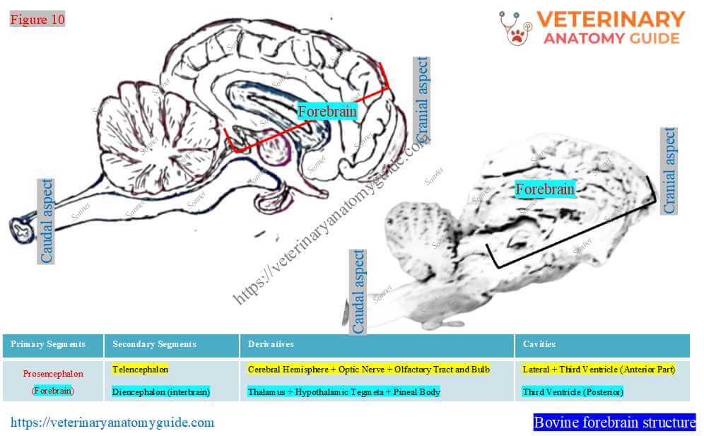
Can you list the anatomical features included under the forebrain segment? Try it yourself, or use Figures 4-7; you will definitely list these features.
All the figures provided here on cow’s brain anatomy are important. But for a description of each part of the cow’s brain, you can always follow Figure 2, followed by Figures 4-7.
Telencephalon (first division of prosencephalon)
According to Figure 2, the first division of the forebrain (prosencephalon) is the telencephalon. However, this part of the forebrain includes the right and left cerebral hemispheres, the optic nerve, and the olfactory tracts and bulbs.
The two lateral ventricles and the anterior part of the third ventricle are also included under the telencephalon.
Here, I will focus only on the features of the cerebral hemisphere, as the other parts need separate descriptions.
Cow’s cerebral hemisphere (cerebrum)
Figure 11 highlights some important features of the cow’s cerebral hemisphere. The falx cerebri of the cranial meninges remains lodged in the longitudinal fissure between the two cerebral hemispheres.
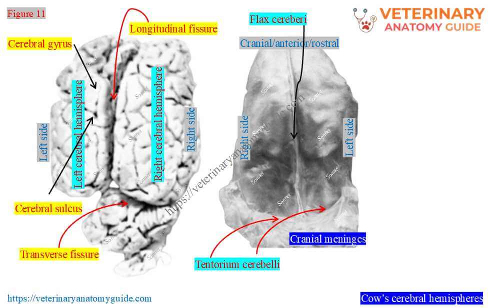
For descriptive purposes, each cerebral hemisphere presents four surfaces, two extremities, and various lobes.
Surfaces of the cerebral hemisphere
Each cerebral hemisphere has four surfaces: dorsal, ventral, lateral, and medial (Figure 12).
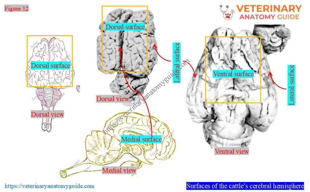
Dorsal surface: This surface is convex and adapted to the roof of the cranial cavity.
Ventral surface: The ventral surface is also known as the basal surface. It presents the piriform lobe, which is located lateral to the cerebral crus and the hypothalamus.
Rostral to the piriform lobe, the basal surface presents a triangular area termed the olfactory trigone. Again, the middle of the basal surface has a deep depression for the middle cerebral artery.
Lateral surface: It is convex and blends with the dorsal surface.
Medial surface: The medial surface of the cerebrum is flat and vertical. It forms the lateral boundary of the longitudinal fissure.
Extremities of the cerebral hemisphere
Here, Figure 13 highlights two extremities of the cerebral hemisphere of the cow’s brain. It shows the rostral and caudal extremities of the cerebral hemisphere.
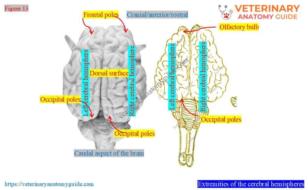
Rostral extremity: Here, the rostral extremity is known as the frontal pole. It is bilaterally compressed, convex, and partially covered by the olfactory bulb.
The olfactory bulb is lodged in the ethmoidal fossa of the cow’s skull. However, it continues caudally and attaches to the cerebral hemisphere by the olfactory tract.
Rostrally, the fibers of the first cranial nerve (olfactory) connect to the olfactory bulb.
Caudal extremity: The caudal extremity of the cerebral hemisphere is also known as the occipital pole. The figure shows that this extremity is wide and arched.
It is flat on its ventromedial aspect and separated from the cerebellum by a transverse groove. Again, this groove is filled with the membranous fold of the tentorium cerebelli.
Lobes of the cerebral hemispheres
The exterior of each cerebral hemisphere of the cow’s brain is thrown into several ridges. These are cerebral gyri, separated by fissures termed sulci.
Figure 14 presents four different lobes of the cerebral hemispheres: frontal, parietal, temporal, and occipital.
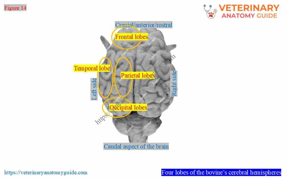
Frontal lobe: It is the most rostral part of the cerebral hemisphere. This lobe controls voluntary muscle movement.
Parietal lobe: It is immediately caudal to the frontal lobe of the cow’s brain. However, the central fissure separates it from the frontal lobe, and the lateral fissure separates it from the temporal lobe.
Temporal lobe: This temporal lobe is ventral to the lateral fissure and rostral to the occipital lobe.
Occipital lobe: It is the most caudal lobe of the cerebral hemisphere. This occipital lobe is associated with vision.
Diencephalon (interbrain) of the cow brain
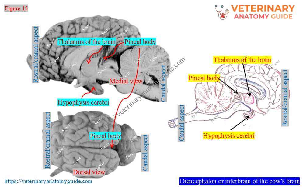
The diencephalon, or interbrain, is the second part of the prosencephalon and consists of the thalamus, hypothalamus, and pineal body. However, it also contains the posterior part of the third ventricle (Figure 15).
Thalamus (thalami) of the brain
The thalamus is a large ovoid mass located obliquely across the dorsal face of the cerebral peduncle (Figure 15). Here, the right and left thalami of the cow’s brain meet at a right angle in front.
Dorsal surface: It is convex and separated from the hippocampus by the tela choroidea.
Lateral part: It is separated from the caudate nucleus by the white-matter band of the stria terminalis.
Posterior part: The posterior part of the thalamus of the cow’s brain is rounded and continues laterally with the optic tract. You will find the medial geniculate body between the thalamus and the cerebral peduncle.
Why is the thalamus important? In a cow’s brain, it acts as the relay and control station between the brainstem and spinal cord.
Hypothalamus and pineal body
The figure also shows the location of the hypothalamus and pineal body in the cow’s brain anatomy. Here, the pineal body is the small fusiform gland in the cow.
However, the cow’s pineal body is located in the depression between the thalamus and corpora quadrigemina.
Cow’s midbrain anatomy (mesencephalon)
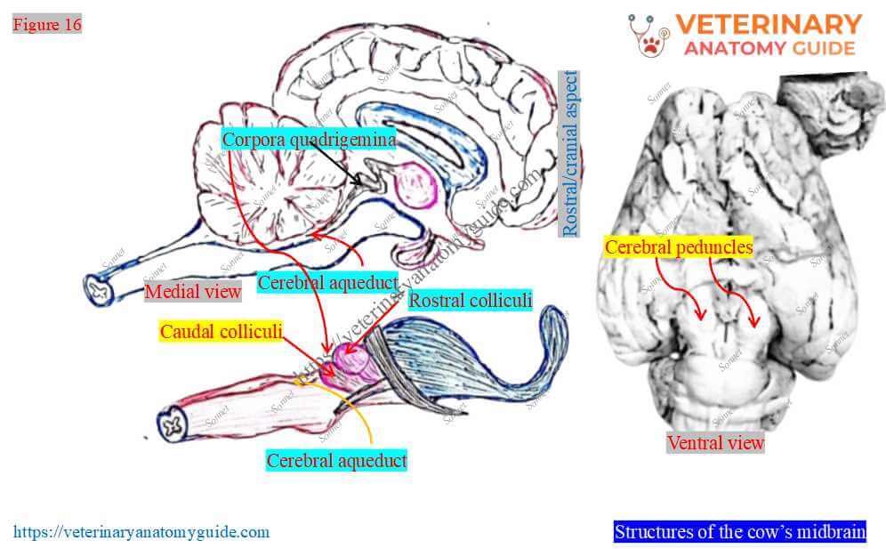
Figure 16 presents all the structures of the cow’s midbrain (mesencephalon). Thus, the midbrain consists of the cerebral peduncle, corpora quadrigemina, and cerebral aqueduct.
Cerebral peduncle of the brain
Figures 2 and 16 identify the cerebral peduncles on the ventral surface of the cow’s brain. These are parts of the midbrain and are two large columns.
Each peduncle emerges from under the pons on either side of the midline. The optic tracts cross the anterior and lateral surfaces of each peduncle.
The medial surfaces of both peduncles form the lateral boundary of the interpeduncular fossa. However, numerous vessels pierce this fossa.
Each of the cerebral peduncles of the cow’s brain divides into the following three parts –
- Dorsal part (tegmentum): It is continuous with the pons and contains some fiber tracts.
- Middle part (substantia niagra): It is a layer of the grey matter and contains various deeply pigmented nerve cells.
- Ventral part (crus cerebri): Here, the ventral part consists of corticospinal, corticopontine, and corticobulbar fibers.
Corpora quadrigemina and cerebral aqueduct
The figure also highlights the corpora quadrigemina and cerebral aqueduct from the cow’s midbrain.
Corpora quadrigemina (tectum/colliculus): These are the two pairs of rounded bodies at the dorsal aspect of the cow’s midbrain. They are known as the paired superior (rostral) and inferior (caudal) colliculi.
Here, these two pairs of colliculi are separated by the shallow transverse groove. The rostral colliculi are grey and lie higher than the caudal colliculi. Here, the caudal colliculi of the cow’s brain are pale and smaller.
The rostral colliculi are separated from the thalamus by a wide transverse groove. Again, the caudal colliculi are also separated from the pons by a transverse furrow.
Cerebral aqueduct: This is the narrow passage in the cow’s mesencephalon (midbrain). It connects the third and fourth ventricles and lies in the median plane.
Hindbrain of the cow (Rhombencephalon)
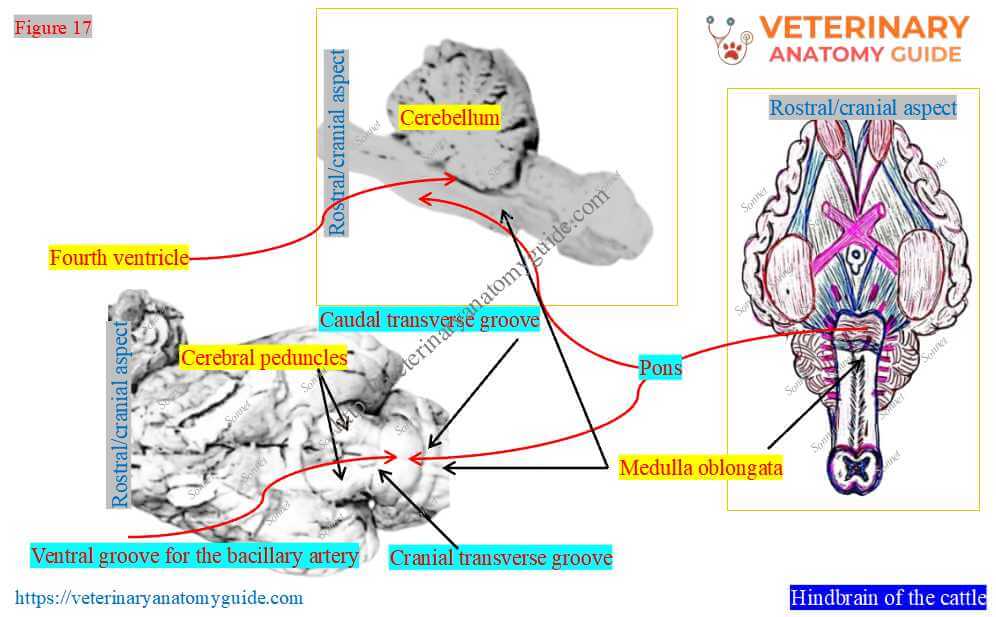
Here, Figure 17 highlights the most caudal part of the cow’s brain anatomy. It is the cow’s hindbrain and consists of the cerebellum, pons, and medulla oblongata.
However, the hindbrain (rhombencephalon) of the cow’s brain divides into two parts (also shown in Figure 2) –
- Metencephalon: It consists of the pons and cerebellum.
- Meyelencephalon: It consists of the medulla oblongata.
However, the fourth ventricle is also included under the hindbrain.
Metencephalon of the cow’s brain
Here, you need to describe the anatomical features of the pons and cerebellum parts of the cow’s brain.
Pons of the cow’s brain
The pons is a transverse protuberance on the ventral surface of the brainstem (Figure 17). However, it lies between the medulla and cerebral peduncles.
The figure shows that the pons is marked off from these structures ventrally by cranial and caudal transverse grooves.
Median ventral groove: The ventral surface of the pons presents the median ventral groove for the basilar artery.
A large part of the pons curves dorsally and continues into the base of the cerebellum. However, the dorsal surface is blended with the rostral cerebral peduncles.
Formation of the fourth ventricle: The central free part of the dorsal surface of the pons forms the rostral part of the fourth ventricle.
Cerebellum of the brain
Figure 18 shows that the cerebellum of the cow’s brain lies caudal to the cerebrum. It overlies the pons, the rostral part of the medulla oblongata, and the fourth ventricle.
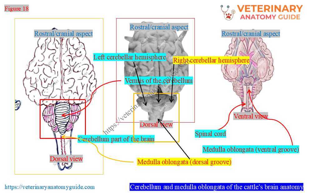
The figure also shows that the cerebellum of the cow/bovine brain is globular and compressed dorsoventrally.
Connection: The cerebellum connects with the medulla, pons, and midbrain via three cerebellar peduncles.
Lobes: It presents three lobes: the median vermis and two lateral cerebellar hemispheres.
Here, the vermis lobe is curved circularly. Its rostral extremity attaches to the rostral medullary velum.
Again, the vermis extends from the caudal aspect of the colliculi to the medulla oblongata. The circular fissures separate the vermis and lateral hemispheres.
The outer surface of the cerebellum of the cow’s brain also shows gyri and sulci, like the cerebrum.
Myelencephalon (medulla oblongata)
Figure 18 also highlights the medulla oblongata of the cow’s brain. It is the cranial continuation of the cow’s spinal cord and forms the caudal part of the brainstem.
However, the cow’s brainstem extends from the foramen magnum to the caudal part of the pons. It is dorsoventrally compressed and presents two surfaces and two extremities.
Surfaces: Both the dorsal and ventral surfaces are convex and present the median fissures. The caudal part of these fissures is faint and becomes deeper.
Extremities: The medulla oblongata is wider at its rostral extremity and narrower toward the caudal extremity.
The medulla oblongata regulates respiration, body temperature, and heart rate. However, the cranial nerves arise from or attach to the medulla oblongata.
FAQ’s on the cow’s brain anatomy
The hippocampus is a small subcortical area of the cow’s brain. However, it is located in the medial temporal lobe of the cow’s cerebral hemisphere (Figure 19).
Again, it is a gyrus that curves from the deep face of the cow’s piriform lobe around the thalamus. The right and left hippocampi connect at their upper part via the hippocampal commissure.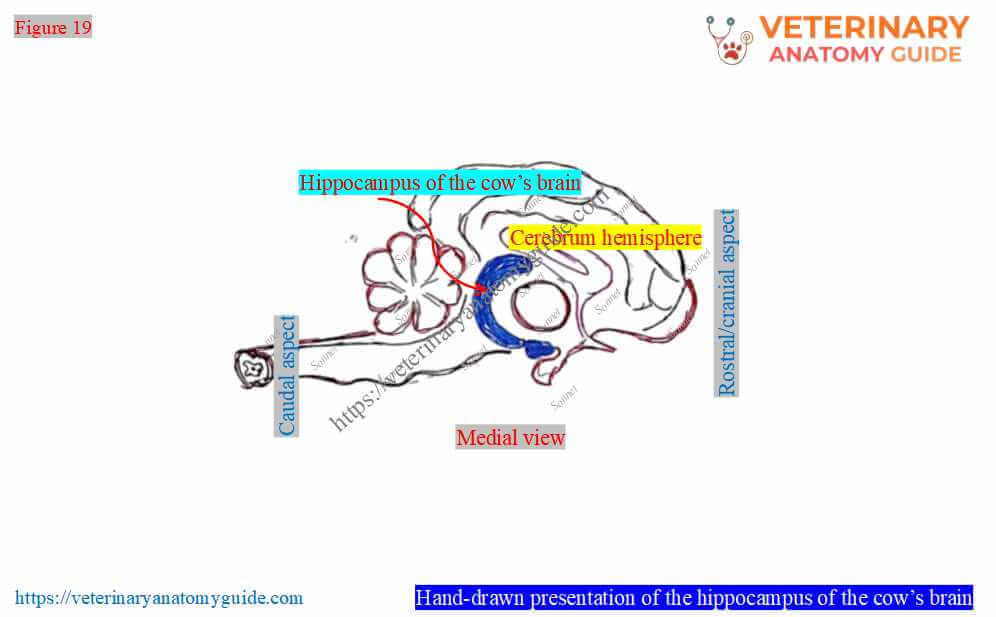
The outer layer of the cerebral hemisphere presents the grey matter (cortex). In contrast, the inner, smaller area is the white matter (medulla).
Here, the grey matter (cortex) is made of nerve cells (neurons). The white matter (medulla) consists of white, myelinated axons of nerve fibers.
Conclusion
Thus, the cow brain anatomy is part of the central nervous system, which is divided into three major divisions. Here, the forebrain mainly contains the cerebrum, thalamus, hypothalamus, and pineal body.
The midbrain of a cow mainly consists of the cerebral peduncles and corpora quadrigemina. Finally, the hindbrain includes the pons, medulla oblongata, and cerebellum segments.
References
- Ballarin, C., et al. (2016). Brain of the Domestic Bos taurus. PloS one, 11(4), e0154580.
- Ghosh, R. K. (2023). Primary veterinary anatomy, 2nd Ed.Current Books International, Kolkata.
- Hermanson et al.: Miller and Evan’s Anatomy of the Dog, 5th Ed. Elsevier.
- Rowen D. Frandson, W. Lee Wilke, and A. Dee: Anatomy and physiology of farm animals, 4th edn.Wiley-Blackwell, USA
- Nomir, A.G., et al. Head of Zebu cattle (Bos Taurus indicus). BMC Vet Res 20, 318 (2024).
- Getty, R., Sission, and Grossman’s The anatomy of the domestic animals, 5th Ed. W. B. Saunders and Co., Philadelphia (USA).
- Schmidt, M. J., et al. (2009). Neuroanatomy of the calf brain. Journal of Morphology, 270(6), 745–758.
- Nickel, R., Schummer, A. and Seiferle, E.: The anatomy of domestic animals, 1st Edn. Verlag Paul Parey, Berlin.
- McLeod, W. N.: Bovine anatomy, 2nd Edn. Burgess Publishing Co. Minneapolis.
- Sibiya, G., et al. (2024) Exploring the anatomical configurations of the cerebral arteries. Sci Rep 14, 6060.
- Dyce, K. M., W. O. Sack, and CSG Wensing: Textbook of Veterinary Anatomy, Lea & Febiger. Philadelphia
Written by