
Animal phrenic nerve anatomy describes its formation, course, and distribution to the thorax and diaphragm. It is a motor nerve (Getty, 1970) to the animal’s diaphragm and comes from the cervical spinal nerves.
In this guide, I will describe the anatomy of the phrenic nerve in animals (cow, sheep, goat) with hand-drawn diagrams and real pictures.
Quick answer: The animal phrenic nerve comes from the ventral primary branches of the last few cervical spinal nerves and innervates the diaphragm.
The formation and distribution (course) of this phrenic nerve in animals (especially in cow, sheep, and goat) is a little complex. However, I will try to present its anatomy simply so you can learn it easily.
How is the animal’s phrenic nerve formed?
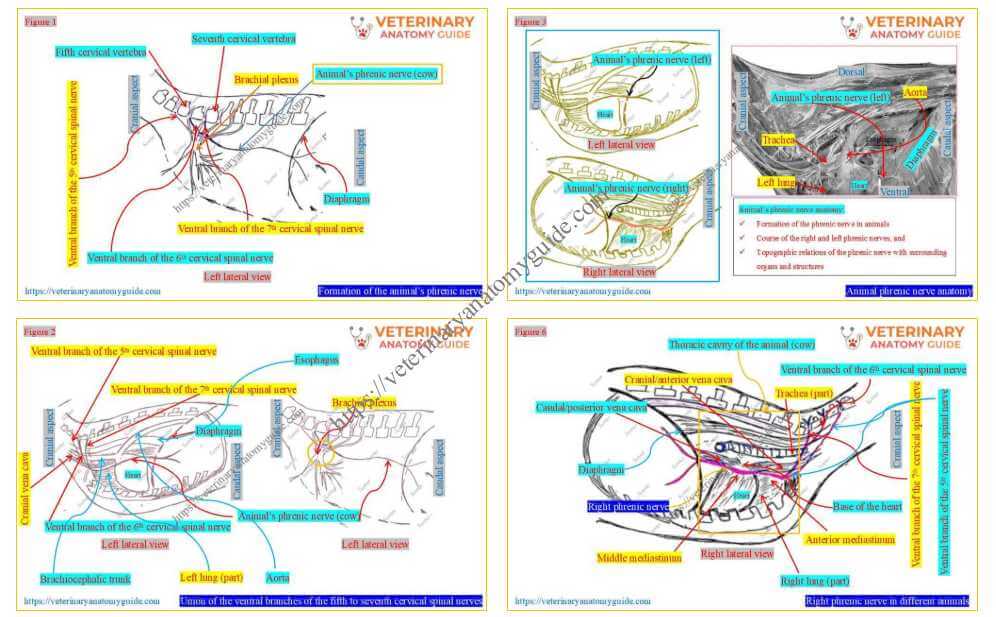
Figure 1 shows the formation of the phrenic nerve in the animals (cow, sheep, goat). It is a hand-drawn picture where I tried to show how the cervical spinal nerves contribute to forming this phrenic nerve.
Prior knowledge of the basic structure of a spinal nerve may help you understand the formation of the animal’s phrenic nerve. Here, the dorsal and ventral roots, and the dorsal and ventral rami before and after the spinal nerve, respectively, are important.
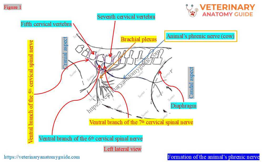
Formation of the phrenic nerve in cow/sheep/goat: The phrenic nerve forms from the union of the ventral branches of the fifth, sixth, and seventh cervical spinal nerves. These branches run obliquely downward and backward over the superficial face of the scaleneus muscle of the thorax.
Sometimes, the ventral branches of the fourth cervical spinal nerve of the animal (cow/sheep) contribute to the formation of this nerve. First, the sixth and seventh nerves unite to form a trunk, and then the fifth nerve joins it within the thoracic cavity (Figure 2).
Thus, the main branches come from the ventral branches of the animal’s sixth and seventh cervical spinal nerves. Phrenic nerve formation differs slightly between large (cow, buffalo) and small ruminants (sheep, goat).
The root (branch) derived from the fifth cervical spinal nerve is small and inconsistent. However, the root from the seventh cervical spinal nerve arises via the brachial plexus.
Animal phrenic nerve anatomy (cow/sheep/goat)
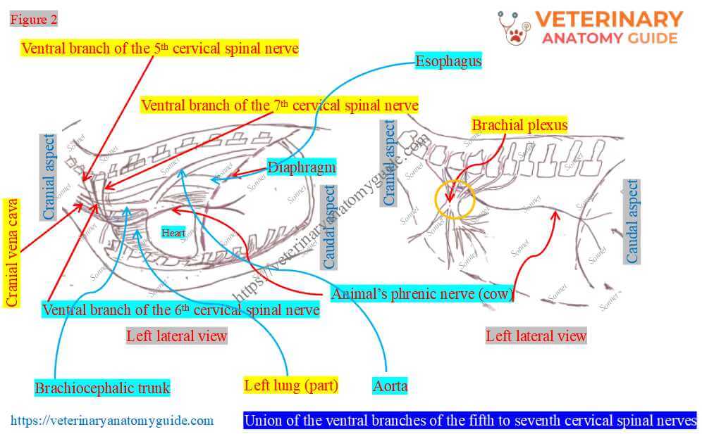
Figure 3 presents an overview of the phrenic nerve anatomy in animals (cow/bovine/sheep/goat). To make it easier, I used the cow’s phrenic nerve anatomy as the ideal description.
This overview and these diagrams might help you retain knowledge of the animal’s phrenic nerve. Thus, you can rely on this guide to prepare for the phrenic nerve written examination.
The figure ensures that the following topics should be mentioned while describing the animal’s phrenic nerve –
- Formation and location of the phrenic nerve
- Course of the animal’s (cow/sheep/goat) phrenic nerve
- Right and left phrenic nerves of the animals, and
- Topographic relations of the organs during their (right and left phrenic) courses
The animal’s phrenic nerve extends from the thoracic inlet to the diaphragm. However, Figures 1 and 2 describe how this nerve forms in animals (cow/goat).
Now, I will describe the course or distribution of the animal’s phrenic nerves with the diagram and real pictures.
How many phrenic nerves are there in animals (cow/sheep/goat)?
Figure 4 clearly indicates the number of phrenic nerves in the animals. Animals have two (right and left) phrenic nerves.
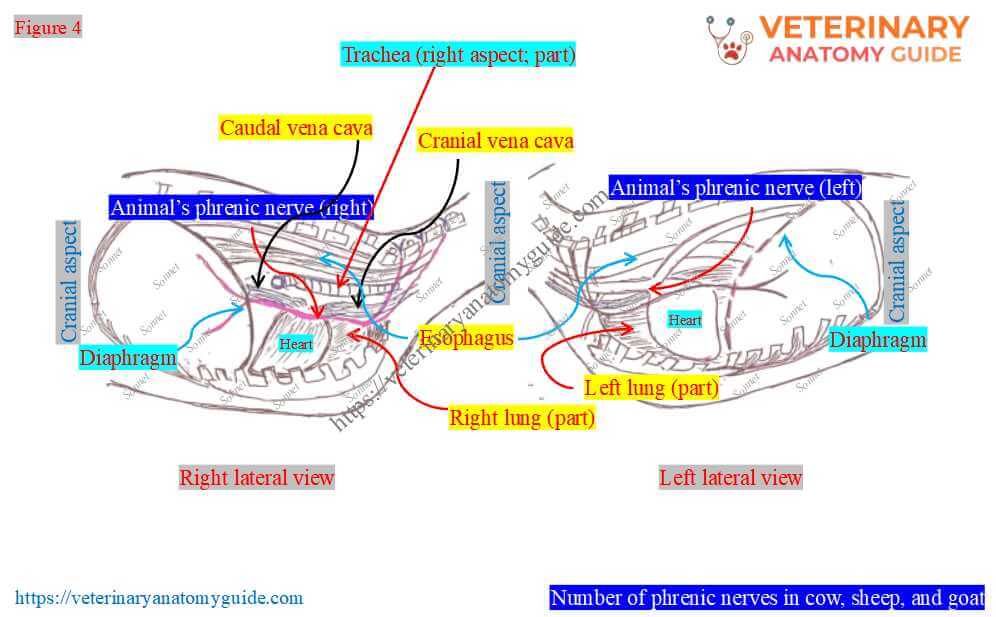
The origin of the right and left phrenic nerves is similar in animals. They cross the lateral surface of the scalenus ventralis and enter the thoracic cavity together.
Course of the animal’s phrenic nerve
Figure 5 provides a schematic (hand-drawn) diagram of the course of the animal’s phrenic nerve. You may follow the phrenic nerve memorization technique shown in the figure.
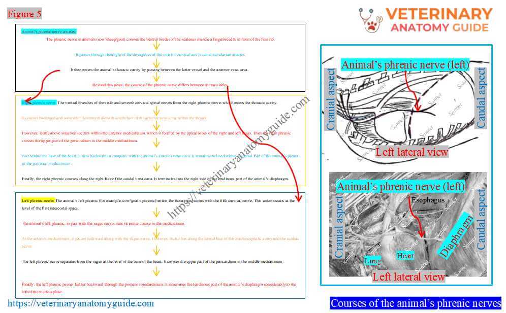
Course: The phrenic nerve in animals (cow/sheep/goat) crosses the ventral border of the scalenus muscle a fingerbreadth in front of the first rib. It passes through the angle of the divergence of the inferior cervical and brachial/subclavian arteries.
It then enters the animal’s thoracic cavity by passing between the latter vessel and the anterior vena cava. Beyond this point, the course of the phrenic nerve differs between the two sides.
Thus, I need to describe the course from this point separately for the right and left phrenic nerves in animals. However, the major course of the right and left phrenic nerves is through the mediastinum of the thorax.
Right phrenic nerve course and topographic relationship in animals
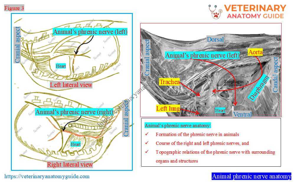
Figure 6 shows an overview of the right phrenic nerve course in the animal (cow/sheep/goat). It also shows the topographic relationship of this nerve with surrounding organs and structures as it courses within the thorax.
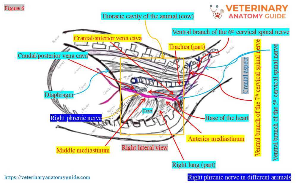
Right phrenic nerve: The ventral branches of the sixth and seventh cervical spinal nerves form the right phrenic nerve, which enters the thoracic cavity. It courses backward and somewhat downward along the right face of the anterior vena cava within the thorax.
However, it (the above situation) occurs within the anterior mediastinum, which is formed by the apical lobes of the right and left lungs. Then the right phrenic crosses the upper part of the pericardium in the middle mediastinum.
Just behind the base of the heart, it runs backward in company with the animal’s anterior vena cava. It remains enclosed within a special fold of the animal’s pleura in the posterior mediastinum.
Finally, the right phrenic courses along the right face of the caudal vena cava. It terminates into the right side of the tendinous part of the animal’s diaphragm.
Course of the left phrenic nerve in animals and topographic relationships
Figure 7 highlights the course of the animal’s left phrenic nerve within the thorax. It also highlights the topographic relations of the left phrenic nerve with the surrounding organs.
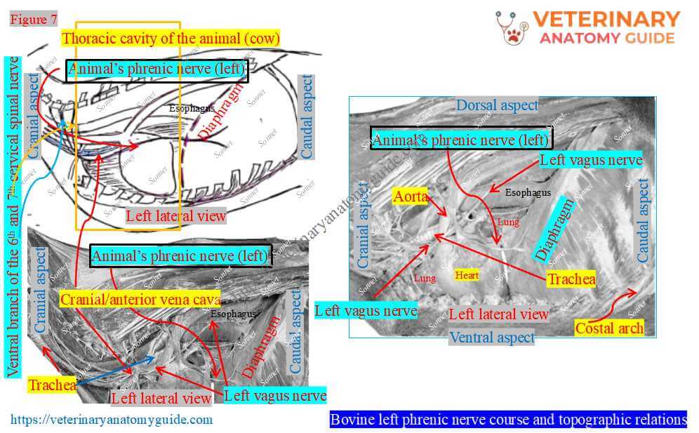
Left phrenic nerve: The animal’s left phrenic (for example, cow/goat’s phrenic) enters the thorax and unites with the fifth cervical nerve. This union occurs at the level of the first intercostal space.
The animal’s left phrenic, in part with the vagus nerve, runs its entire course in the mediastinum.
At the anterior mediastinum, it passes backward along with the vagus nerve. However, it also lies along the lateral face of the brachiocephalic artery and the cardiac nerve.
The left phrenic nerve separates from the vagus at the level of the base of the heart. It crosses the upper part of the pericardium in the middle mediastinum.
Finally, the left phrenic passes further backward through the posterior mediastinum. It innervates the tendinous part of the animal’s diaphragm considerably to the left of the median plane.
The animal’s phrenic nerves are usually connected near the origin to the first thoracic sympathetic ganglion. Again, each terminates by dividing into several branches which are distributed to the corresponding part of the diaphragm.
FAQ’s on animals’ phrenic nerves
The fifth to seventh ventral branches of the cervical spinal nerves enter the thorax and unite to form the cow’s/cattle phrenic nerves. These nerves innervate the diaphragm to maintain respiration; thus, they are unique.
The animal’s cervical spinal nerves are eight pairs and pass through the corresponding intervertebral foramen, except the first two. Here, the first cervical spinal nerve leaves the spinal canal through the lateral vertebral foramen of the atlas vertebra.
Again, the second cervical spinal nerve emerges from the canal through the lateral vertebral foramen of the axis bone.
Conclusion
Thus, the phrenic nerve of the animal arises from the ventral branches of the fifth to seventh cervical nerves. This nerve mainly courses through the animal’s mediastinum.
However, animals (cow, sheep, goat) have two phrenic nerves: right and left. They course differently after their origin and innervate the corresponding tendinous part of the animal’s diaphragm.
References
- Bolat, D., S. et al., 2026. Anatomy of the Phrenic Nerve in Sheep. Anato. Histo. Embryo. 55 (4): e70139.
- Rowen D. Frandson, W. Lee Wilke, and A. Dee: Anatomy and physiology of farm animals, 4th edn.Wiley-Blackwell, USA.
- Dos Anjos , C.S. É. A., et al. (2025). Origin and Distribution of the Phrenic Nerve in the Diaphragm. Anatomia, histologia, embryologia, 54(4), e70046.
- Getty, R. Sission and Grossman’s The anatomy of the domestic animals, 5th Edn. W. B. Saunders and Co., Philadelphia (USA).
- Nickel, R., Schummer, A. and Seiferle, E.: The anatomy of domestic animals, 1st Edn. Verlag Paul Parey, Berlin.
- Ghosh, R. K.: Primary Veterinary Anatomy, 2nd Ed.Current Books International, Kolkata.
- Hermanson et al.: Miller and Evan’s Anatomy of the Dog, 5th Ed. Elsevier.
- McLeod, W. N.: Bovine Anatomy, 2nd Ed. Burgess Publishing Co., Minneapolis.
- Dyce, K. M., W. O. Sack, and CSG Wensing: Textbook of Veterinary Anatomy, Lea & Febiger. Philadelphia
- Byers, S., et al. (2011). Neurological causes of diaphragmatic paralysis in 11 alpacas (Vicugna pacos). J Vet Intern Med, Vol. 25:380–385.
Written by