
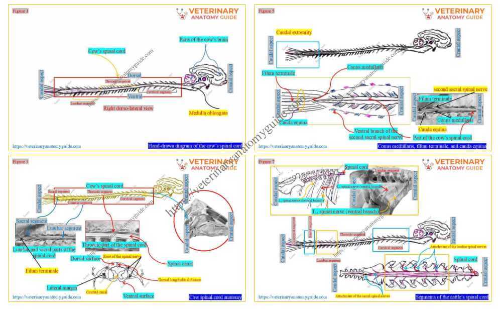
Cow spinal cord is an important connector between the brain and the body. However, it is the caudal continuation of the medulla oblongata, part of the brainstem.
In this guide, I will describe the anatomical features of the cow’s spinal cord with hand-drawn diagrams and real pictures.
Quick answer: The cow spinal cord is a thick, cylindrical structure located in the vertebral canal that extends from the foramen magnum to the middle of the sacrum.
Here, I will help you understand the formation and extension of the cow’s spinal cord in a practical way. However, I will also describe its internal structure, which consists of inner grey and outer white matter.
What is the cow’s spinal cord?
Figure 1 presents a hand-drawn presentation of the cow’s spinal cord. The figure also shows that it links the brain to the peripheral nerves of the cow’s body.
Thus, the spinal cord is part of the cow’s central nervous system, where a mass of nervous tissue is arranged in a cylindrical form.
Location of the cow’s spinal cord
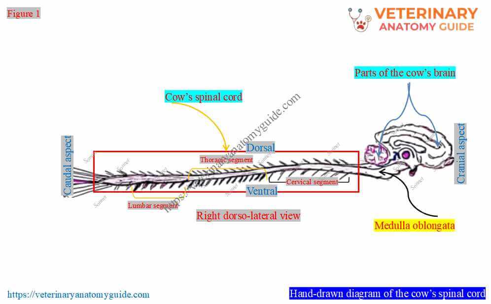
Figure 2 highlights the location of the cow’s (bovine) spinal cord. It shows the starting and ending points of the spinal cord with some peculiar features.
Location: According to the figure, the cow’s spinal cord lies within the vertebral canal, formed by various segments of the vertebrae. However, a strong covering called the spinal meninges loosely covers the spinal cord within the canal.
The above figure also helps you to understand the actual extension of the bovine/cow’s spinal cord.
Extension: Thus, the spinal cord extends from the level of the cow’s foramen magnum of the occipital bone of the skull, or the medulla oblongata. However, it ends at the level of the cow’s second sacral vertebra (Figure 2).
Cow spinal cord anatomy
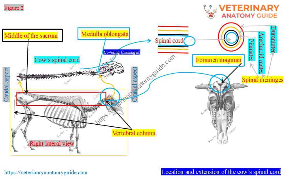
Figure 3 shows an overview of the cow’s spinal cord anatomy. It shows the cord’s appearance (diameter/thickness, surfaces, margins, and extremities), its coverings, and its attachment to the spinal nerves.
Thus, from this figure, you can understand what features you should cover for the cow’s spinal cord anatomy –
- Location and extension of the cow’s spinal cord
- Diameter and extremities of the spinal cord
- Surfaces of the bovine’s spinal cord, and
- Segments and attachment of the spinal nerves to the spinal cord
Figures 1 and 2 describe the location and extension of the bovine spinal cord. Thus, I will start with the diameter and extremities of the spinal cord.
However, I suggest that you write the anatomy of the cow’s spinal cord accordingly in your examination paper, with proper diagrams.
Diameter and extremities of the bovine spinal cord
Figure 4 shows changes in the diameter, or thickness, of the cylindrical spinal cord. It shows that the spinal cord is not uniform in its diameter throughout its length.
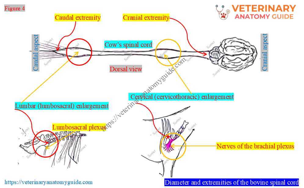
Diameters: As the cow’s spinal cord is continuous with the medulla oblongata, its beginning is dilated. However, the figure shows two other dilatations at the level of the origin of the nerves of the cow’s brachial plexus and the lumbosacral plexus.
Again, these dilatations are known as the cervical (cervicothoracic) and lumbar (lumbosacral) enlargements. Here, the caudal extremity of the spinal cord gradually diminishes in volume from the lumbar region.
What are the conus medullaris, filum terminale, and cauda equina?
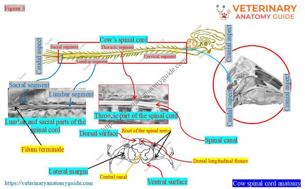
Figure 5 highlights the caudal extremity of the cow’s spinal cord. It shows three structures: the conus medullaris, filum terminale, and cauda equina.
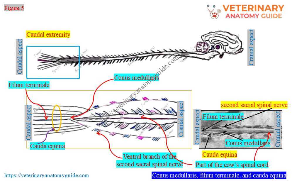
Let me explain these terms or structures of the spinal cord perfectly:
- Conus medullaris: The figure shows that the caudal part of the cow’s spinal cord terminates in a tapering and pointed manner. It occurs at the level of the cow’s second sacral vertebra. This pointed terminal part of the cow’s spinal cord is known as the conus medullaris.
- Filum terminale: The figure also shows that a very thin structure extends from the conus medullaris to the first coccygeal vertebra. This thin spinal cord structure is termed the filum terminale.
- Cauda equina: A lash of long nerve roots of some sacral and coccygeal spinal nerves occupies the caudal part of the vertebral canal. However, these roots attach to the spinal cord and, as a whole, look like the tail of a horse, which is termed the cauda equina.
Here, the filum terminale of the cow’s spinal cord is not a nervous element. Instead, it is a slender filament of the pia mater.
Surfaces of the bovine or cow’s spinal cord
Figure 6 presents the two surfaces of the cylindrical spinal cord of a cow (bovine/cattle). Although the spinal cord is cylindrical, the figure shows it is slightly dorsoventrally compressed.
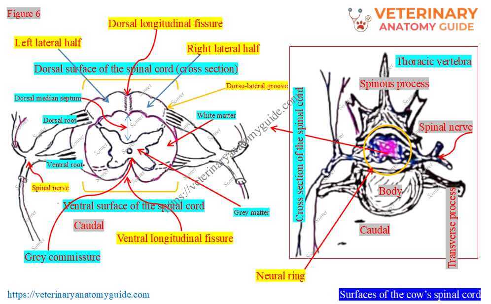
The figure also shows that the dorsal and ventral surfaces of the spinal cord present longitudinal median fissures. However, these fissures divide each surface into two symmetrical halves: right lateral and left lateral.
Dorsal surface and longitudinal fissure: Here, the dorsal surface is convex and the dorsal longitudinal median fissure is shallow. However, the dorsal median septum fills this fissure.
Ventral surface and longitudinal fissure: The ventral surface of the spinal cord is also convex. However, the ventral longitudinal median groove is narrow and deep, and reaches about the middle of the spinal cord.
Again, the ventral longitudinal fissure accommodates the ventral spinal artery of the cow. The commissures of the grey and white matter connect the two halves of the spinal cord.
Unique features of the dorsal surface: Each dorso-lateral surface of the spinal cord presents a faint groove along its length. These faint grooves (right and left) are the dorsolateral grooves.
However, the ventral surface of the cord has no such ventrolateral grooves.
Segments and attachment of the spinal nerves to the cattle’s spinal cord
Figure 7 presents the five different segments of the cattle’s spinal cord. It also shows how the spinal nerve roots attach to the cord.
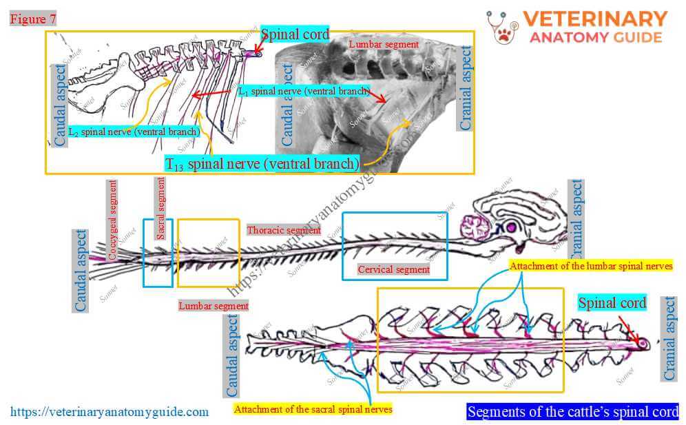
Cows have 37 pairs of spinal nerves that arise from the lateral margin of the spinal cord. Again, these nerves are classified as eight cervical, thirteen thoracic, six lumbar, five sacral, and five coccygeal spinal nerves.
Based on how these nerves attach to the spinal cord, the spinal cord is considered to have five segments. Thus, the segments of the cow’s spinal cord are cervical, thoracic, lumbar, sacral, and coccygeal.
Compared to the vertebral formula of a cow, the spinal nerves are fewer in number. Thus, the cow’s spinal cord and vertebral column grow unequally.
Each spinal nerve is attached to the bovine’s spinal cord by the longer dorsal and short ventral roots. Here, the dorsal root enters the spinal cord at the level of the dorsal lateral groove on the dorsal surface.
Again, the ventral root leaves the spinal cord slightly lateral to the ventral median fissure or groove.
Structure of the cow’s spinal cord
Figure 8 presents an overview of the cow’s spinal cord structure. However, it is a transverse section of the spinal cord that shows the peripheral white matter and inner grey matter.
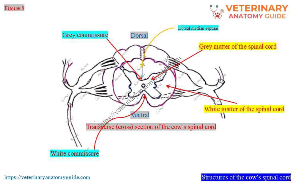
The figure also shows that the white and grey matter appear like the letter H or a butterfly. Here, two wings of the butterfly are connected across the midline by the dorsal grey commissure and, below it, the white commissure.
What is the central canal in the bovine’s spinal cord?
Figure 9 shows a minute canal at the center of the spinal cord and within the grey matter. This is the central canal of the bovine’s spinal cord.
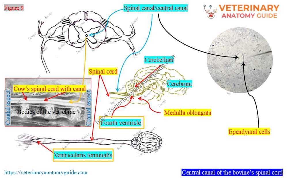
This minute central canal (spinal canal) is filled with cerebrospinal fluid at the center. However, a single layer of tall columnar cells surrounds this central canal.
These columnar cells are termed the ependymal cells of the spinal cord. Again, these ependymal cells are clearly visible under microscopic examination of a transverse (cross-sectional) section of the bovine spinal cord.
Opening of the central canal: The central canal opens rostrally into the caudal angle of the cow’s fourth ventricle. However, it ends at the conus medullaris, forming a slight dilation, termed the ventricularis terminalis.
Grey matter of the cattle’s spinal cord
Figure 10 presents the anatomical features of the grey matter of the cattle’s spinal cord. It mainly contains neuron cell bodies and neuroglia.
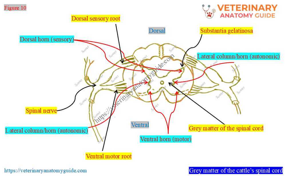
Here, the above figure shows each wing of the grey matter of the cattle’s spinal cord, following 4 columns –
- Dorsal horn (sensory): It consists of the sensory nuclei that receive and process the incoming somatosensory information. From here, the ascending, or sensory, projection of the spinal nerve transmits sensory information to the midbrain and diencephalon.
- Intermediate column (autonomic): It consists of autonomic neurons innervating the visceral organs of the abdominal and pelvic cavities.
- Lateral column/horn (autonomic): This horn is located at the lateral aspect of the grey matter and found only in the thoracic and cranial part of the lumbar regions. However, it is considered the relay station for autonomic sensations, like the intermediate column.
- Ventral horn (motor): Here, the ventral horn of the spinal cord comprises the motor neurons that innervate the skeletal muscles of the cow.
If you look at Figure 10 again, you will see the pointed end of the dorsal horns. They are translucent and called the substantia gelatinosa.
White matter of the bovine’s spinal cord
Figure 11 shows the unique gross features of the white matter of the bovine’s spinal cord.
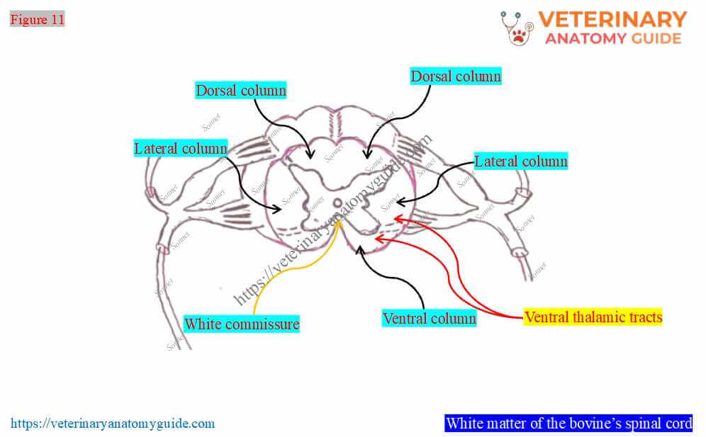
Here, the white matter of the spinal cord consists of dorsal ascending (sensory) and ventral descending (motor) tracts. However, tracts are bundles of fibers that carry one or more sensory or motor impulses in the cow’s central nervous system.
Here, the white matter of the spinal cord divides into the following three pairs of columns –
- Dorsal column: It lies on either side of the dorsal median septum of the spinal cord. However, this column is laterally limited by the dorsal lateral groove.
- Ventral column: It lies on either side of the ventral median septum of the cord. Again, it is limited laterally by the ventral root fibers.
- Lateral column: It lies laterally between the dorsal and ventral columns.
The right and left columns of the spinal cord are connected through the white commissure. However, the white commissure is located outside the grey commissure of the cow’s spinal cord.
FAQ’s on the cow’s spinal cord anatomy
The spinal cord runs caudally from the medulla oblongata without any natural line of demarcation. However, you can identify the actual starting point of the cow’s spinal cord in a real sample.
You will find the opening of the minute spinal canal after the medulla oblongata (Figure 12). This is the actual starting point of the cow’s spinal cord.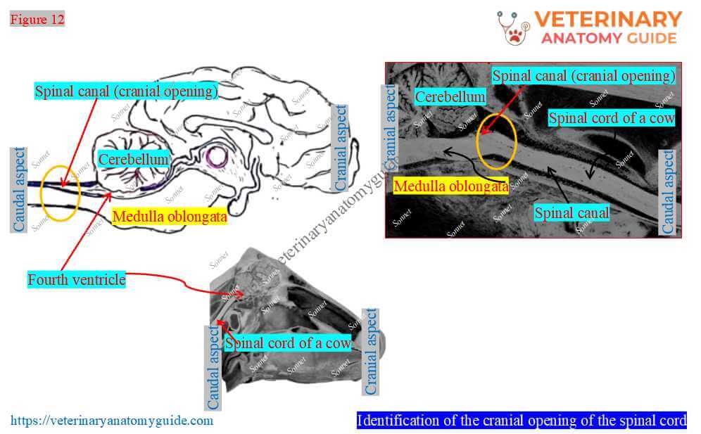
The dorsal column of the white matter in the cow’s spinal cord contains a few important sensory tracts. These sensory tracts are the fasciculus gracilis, fasciculus cuneatus, and common tract of the dorsal column.
Conclusion
Thus, the cow spinal cord is a mass of nervous tissue arranged in a cylindrical form. However, it is enclosed inside the vertebral canal and extends from the medulla oblongata to the middle of the sacrum.
The dorsal and ventral surfaces of the cow’s spinal cord are divided into two halves by the respective median fissures. However, the spinal cord has various horns or columns in both the grey and white matter.
References
- Rowen D. Frandson, W. Lee Wilke, and A. Dee: Anatomy and physiology of farm animals, 4th edn.Wiley-Blackwell, USA.
- Braun, U., et al. (2015). Anatomy of the spinal cord and collection of cerebrospinal fluid from the atlanto-occipital space in cattle. BMC Veterinary Research, 11, 227.
- Ghosh, R. K.: Primary Veterinary Anatomy, 2nd Ed.Current Books International, Kolkata.
- Getty, R. Sission and Grossman’s The Anatomy of the Domestic Animals, 5th Ed. W. B. Saunders and Co., Philadelphia (USA).
- Sokulskyi, I. M., et al. (2021). Histostructure of the gray matter of cattle’s spinal cord. Ukr. Jr. of Vet. and Agri. Sci., 4(3), 11–15.
- Nickel, R., Schummer, A. and Seiferle, E.: The anatomy of domestic animals, 1st Edn. Verlag Paul Parey, Berlin.
- Bassuino, D. M., et al. (2016). Anatomy of spinal cord in cattle and horses. American Association of Veterinary Laboratory Diagnosticians, Inc., 28(4), 455–460.
- Hermanson et al., Miller and Evan’s Anatomy of the dog, 5th Edn. Elsevier.
- Thron, A., et al. Development of the Arterial Supply of the Spinal Cord and Histological Studies in Cattle. Clin Neuroradiol 32, 325–343 (2022).
- McLeod, W. N.: Bovine anatomy, 2nd Edn. Burgess Publishing Co., Minneapolis.
- Dyce, K. M., W. O. Sack, and CSG Wensing: Textbook of Veterinary Anatomy, Lea & Febiger. Philadelphia
- Maya S. (2024). Gray Matter of Spinal Cord in Animals. Ind. Jr. of Ani. Res. 58(10): 1633-1641.
Written by