
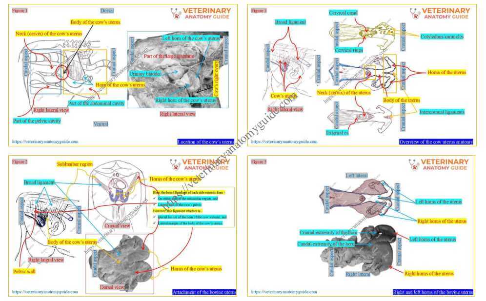
Cow uterus anatomy describes its location, horns, body, and neck, along with the broad ligament. It is a hollow, musculomembranous organ that is continuous with the cow’s uterine tube cranially.
Here, I will describe the detailed anatomy of the cow’s uterus with hand-drawn diagrams and real images.
Quick answer: Cow uterus anatomy includes the horns, body, neck, and the ligament that attaches it to the sublumbar region and pelvic wall.
Thus, I will help you identify the features of the three parts of the cow’s uterus in practice. However, I will also help you understand how the broad ligament keeps the cow’s uterus in position.
Where is the cow’s uterus located?
Figure 1 shows the location of the cow’s uterus in the abdominal and pelvic cavities. However, it is a hand-drawn presentation of the cow’s uterus that also shows its attachment to surrounding structures.
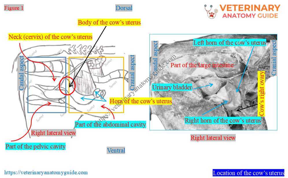
Location: Thus, according to the figure, the cow’s uterus is chiefly located in the caudal part of the abdominal cavity. However, a small part is also located in the cow’s pelvic cavity.
Again, within the pelvic cavity, the cow’s uterus remains between the urinary bladder and parts of the large intestine.
Attachments of the broad ligament to the cow/bovine uterus
Figure 2 highlights the attachment of the cow (bovine) uterus within the abdominal and pelvic regions.
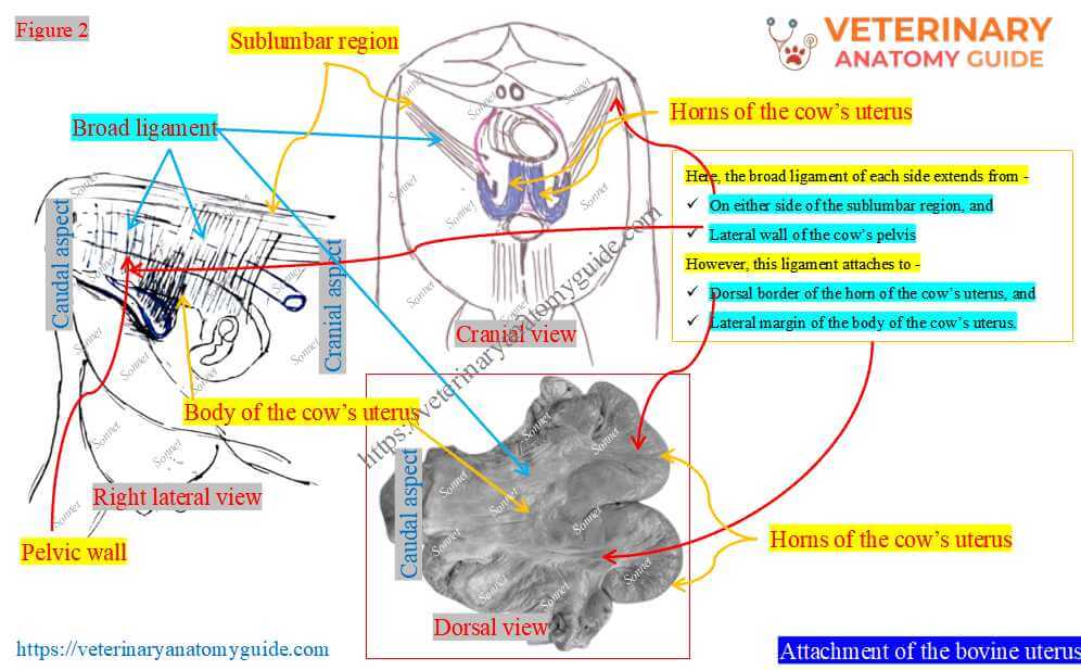
Here, the sublumbar region is formed by the sublumbar muscles under the cow’s lumbar vertebrae. Again, the cow’s pelvic bones along with the surrounding muscles and ligaments form the pelvic region.
Attachment: Thus, the cow’s uterus is attached to the sublumbar areas and lateral walls of the pelvic cavity by two folds of peritoneum. Here, these folds of the cow’s peritoneum are termed the broad ligament.
In the above figure, I also tried to explain the extension of the broad ligament clearly. You can use this to identify variations in the broad ligament’s attachment to the cow’s uterus.
Here, the broad ligament of each side extends from –
- On either side of the sublumbar region, and
- Lateral wall of the cow’s pelvis
However, this ligament attaches to –
- Dorsal border of the horn of the cow’s uterus, and
- Lateral margin of the body of the cow’s uterus.
Now you will find the attachment variation of the broad ligament to the two parts of the cow’s uterus practically.
What is the round ligament of the cow’s uterus?
The lateral layer of each broad ligament gives off a fold to the cow’s uterus. It extends to the internal inguinal ring and is termed the round ligament of the cow’s uterus.
However, this round ligament of the cow’s uterus blends with the parietal peritoneum. Its cranial extremity attaches to the cranial end of the horn of the cow’s uterus.
Cow uterus anatomy
Figure 3 shows an overview of the anatomy of the cow’s uterus. It presents the most important and unique features of the various parts of the cow’s uterus.
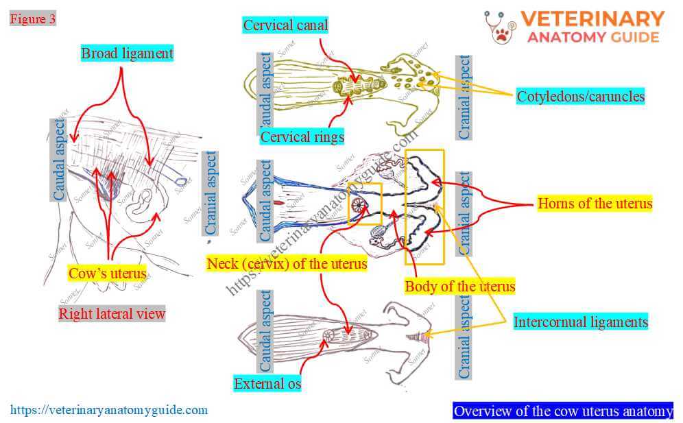
According to the figure, you should describe the following while writing the anatomy of the cow’s uterus –
- Location and attachment of the cow’s uterus,
- Parts of the cow’s uterus along with their unique anatomical features,
- Topographic anatomy of the uterus (I will describe it along with the description of the individual parts of the uterus), and
- Position of the intercornual ligament (You may mention it while describing the horns of the cow’s uterus).
As the first topic (location and attachment) is already completed, I will start with the parts of the cow’s uterus.
Parts of the cow or bovine uterus
Figure 4 highlights the three parts of the cow (bovine) uterus. The first part is the paired horns, or cornua; the middle part is the body; and the caudal part is the neck, or cervix.
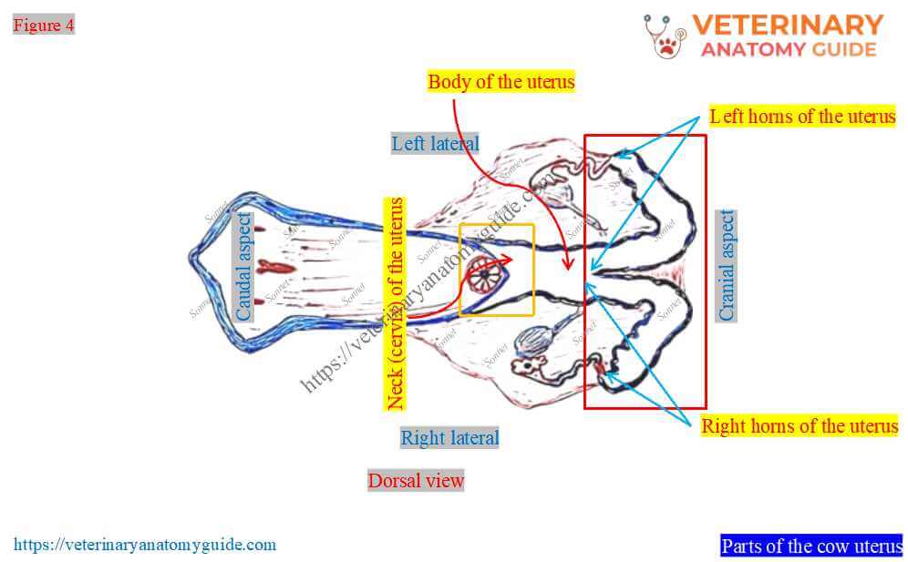
Now, you need to learn the specific and differentiating anatomical features of these three parts of the uterus. However, you might also write these features of the horns, body, and neck in your examination paper.
Horns of the cow’s uterus
Figure 5 shows the right and left horns of the cow’s uterus. Each uterine horn is a cylindrical, spiral muscular tube.
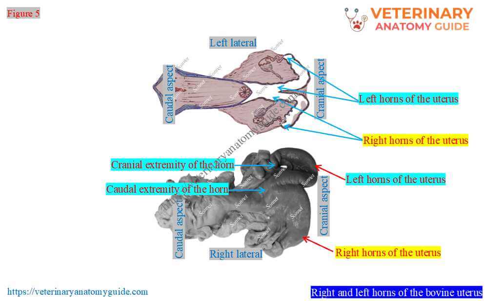
Extremities: These right and left horns of the cow’s uterus present two extremities: cranial and caudal. Here, the cranial extremities of these horns are tapered and join the fallopian tube.
However, the caudal extremities of these horns are wider. Here, the caudal extremities of the two horns unite caudally and form the body of the uterus.
Location and direction of the horns of the cow’s uterus
The horns of the cow’s uterus are located in the abdominal cavity. However, these horns are directed ventrally, cranially, outward, caudally, and upward.
Here, Figure 6 shows the location of the horns of the cow’s uterus. However, this is a hand-drawn presentation where I tried to show the directions of the horns.
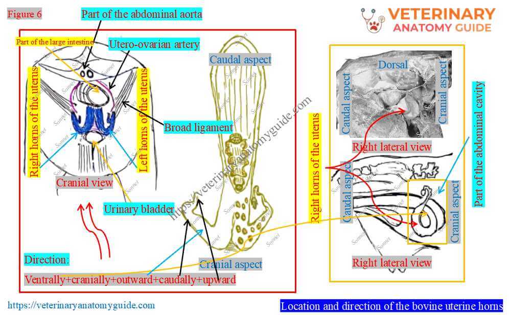
Thus, horn direction is important for understanding horn position and for future palpation of the ovary. However, the broad ligament maintains this spiral arrangement of the horns of the cow’s uterus.
Here, the dorsal border of the horns of the bovine’s uterus is slightly concave. Again, the ventral border is convex and remains free towards the cow’s abdominal cavity.
Why is it named as horns: The spiral appearance of the horns of the cow’s uterus is like the horn of the ram. Thus, this part of the cow’s uterus is termed the horns or cornua.
Length of the horns of the bovine uterus
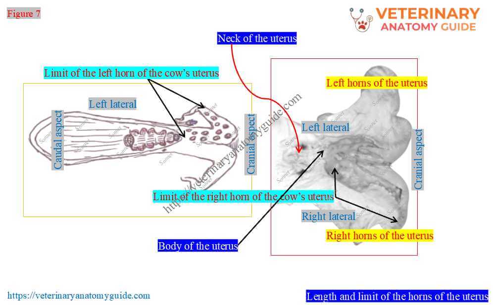
The length of the horns of the uterus is variable, about 35-40 centimeters. However, they appear to be shorter than this length in the practical sample.
In my experience, most students in my gross anatomy classes can’t identify the horns’ length perfectly at first sight. This is because the two horns of the uterus extend cranially and laterally from the body and are united for a short distance.
Again, connective tissue and a common peritoneal layer cover the horns and the body of the cow’s uterus. Thus, it seems to be shorter than the actual length of the horns.
However, the dissected sample of the horns of the bovine uterus can show the actual length and limit (Figure 7). It shows that these two horns diverge and unite at their base to form the body part.
What is the intercornual ligament in the cow’s uterus?
Figure 8 shows the intercornual ligament between the horns of the cow’s uterus.
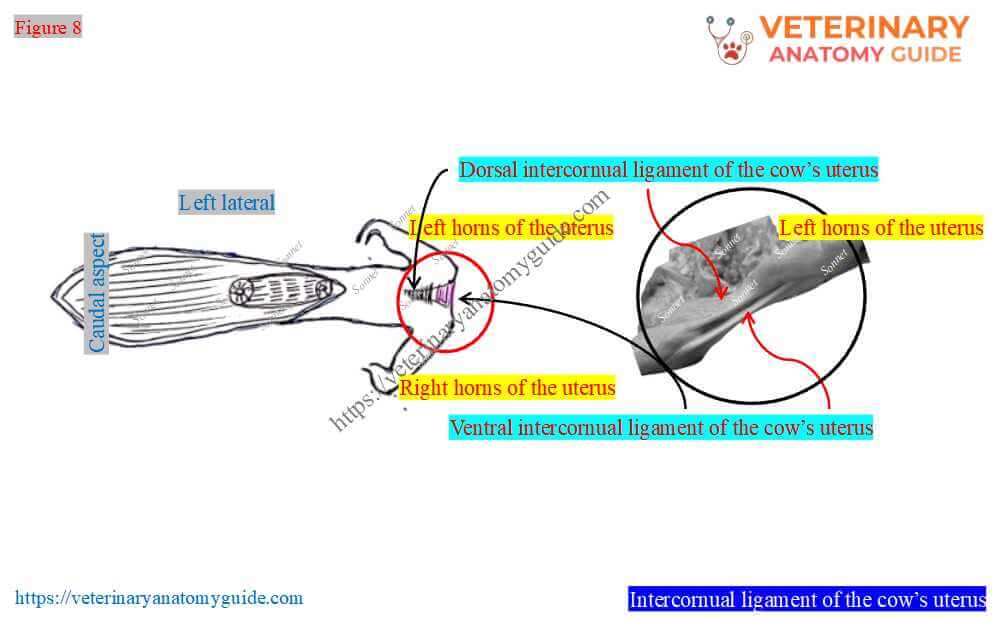
The figure shows that a groove dorsally marks the divergence between the right and left horns of the uterus. This is the intercornual groove, which deepens cranially.
Here, the dorsal and ventral short of the peritoneal folds attach the two horns along the intercornual groove. These structures are termed the intercornual ligaments of the cow’s uterus.
Body of the bovine/cow uterus
Figure 9 highlights the limits and features of the anatomy of the body of the cow uterus. It is a short, cylindrical, dorsoventrally flattened, tube-like structure.
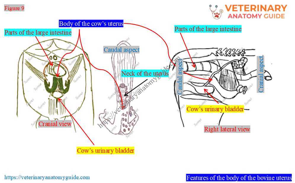
Here, the wall of the body of the cow’s uterus is thicker than the horns. It cranially bifurcates to form the right and left horns. However, caudally, it continues as the neck (cervix) of the cow’s uterus.
Topographic location of the body of the uterus
The body of the cow’s uterus is partly situated in the abdominal cavity and partly in the pelvic cavity. However, the broad ligament attaches to the lateral margin of the uterine body.
The body of the uterus has relations with different organs dorsally and ventrally –
Dorsally: The body is dorsally related to the last two parts of the cow’s large intestine.
Ventrally: It is ventrally related to the urinary bladder and various parts of the cow’s small intestine.
Length and diameters of the body of the cattle uterus
The body of the uterus is actually about 3-4 centimeters long. However, externally it appears to be about 10-12 centimeters long, which is longer than its actual length.
As you already know, this is due to the presence of common peritoneal covering on the caudal part of the horns before their union. However, uterine body length may vary among cows.
Neck (cervix) of the bovine uterus
Figure 10 highlights the neck (cervix) of the cow’s uterus. It is the constricted, caudal-most part of the cow’s uterus.
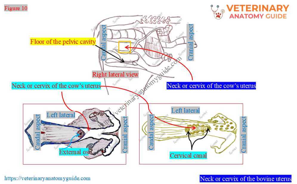
Location: The figure shows that the neck of the cow’s uterus lies on the cranial part of the pelvic floor.
Length and diameter of the neck: It measures about 10 centimeters in length and is remarkably firm and dense in its consistency. However, the external diameter of the uterine neck is almost the same as the diameter of the body.
However, the lumen of the uterine neck is smaller than that of the body. This is due to the excessively thick and firm wall of the neck (cervix) of the cow’s uterus.
The wall of the neck is about 3 centimeters thick. Here, the muscles of the wall of the neck form four annular folds, or rings.
However, these folds or rings are arranged eccentrically. They are visible through the longitudinal dissection of the neck part of the cow’s uterus.
Internal unique anatomical features of the uterine neck
Figure 11 shows the unique internal anatomical features of the neck (cervix) part of the cow’s uterus. Here, the lumen of the neck becomes a narrow canal and remains filled by the spiral folds of the mucous membrane.
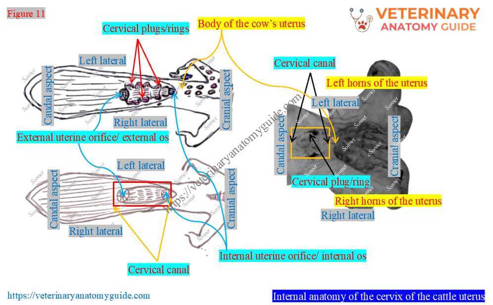
In this area (neck of the uterus), you will find the following important structures –
- Cervical canal/birth canal: This is the narrow canal or passage within the neck part of the cow’s uterus. This passage is spiral because the cervical rings are arranged eccentrically.
- Longitudinal folds and spiral cervical rings: It has various longitudinal mucosal folds and eccentrically located spiral cervical rings. However, the cervical rings are more pronounced in cows than in sheep and goats.
- External uterine orifice (ostium uteri externus/external os): The caudal opening of the cervical canal of the cow’s uterus.
- Internal uterine orifice (ostium uteri internus/ internal os): The cranial opening of the cervical canal into the cavity of the body of the cow’s uterus.
Structure of the cow uterus
Figure 12 presents an overview of the structure of the cow’s uterus. However, it also shows special features of the inner layer of the horns of the cow’s uterus.
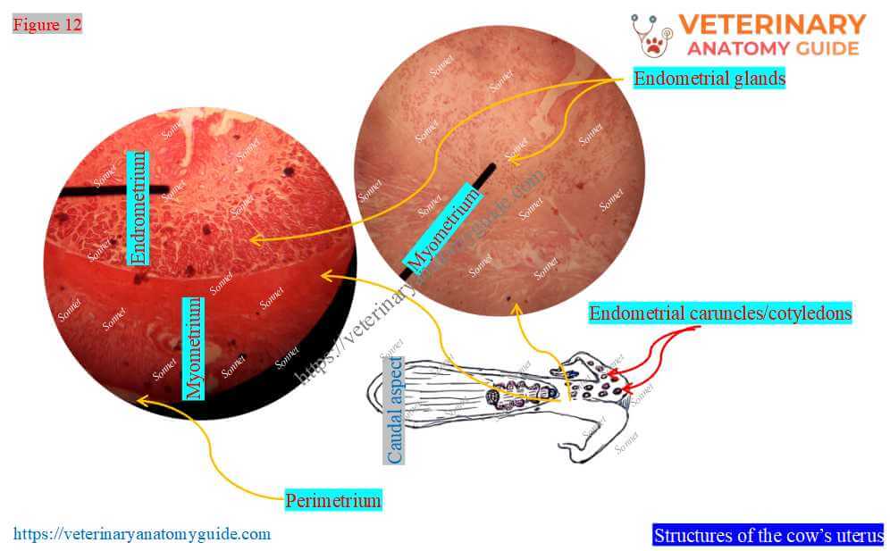
Here, the wall of the uterus consists of the following layers –
- Perimetrium: The outer serous layer.
- Myometrium: This is the middle muscular layer of the cow’s uterus.
- Endometrium: The inner mucous coat of the uterus.
Here, the perimetrium is derived from the peritoneum and covers the entire organ except the lateral margin. However, the myometrium is thick and consists of three layers of smooth muscle.
The endometrium of the cow’s uterus consists of surface epithelium and lamina propria. It shows the coiled and branched endometrial glands that open onto the endometrial surface.
What are endometrial caruncles, or cotyledons, in the cow’s uterus?
Figure 13 highlights the large number of raised, pitted, button-like structures on the endometrial wall of the horns. These structures are scattered and are termed endometrial caruncles or cotyledons.
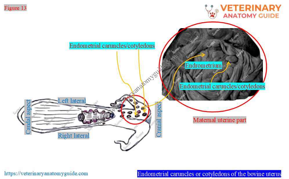
The caruncles of the cow’s uterus are large oval prominences at the middle of the horns. However, they are smaller oval prominences at the end of the horns of the cow’s uterus.
Here, the crypts of the cotyledon or caruncles receive the chorionic villi of the cow’s placenta. Uterine vessels enter the caruncles through the surface attached to the uterine wall.
Vessels and nerves of the bovine uterus
Arteries: The cranial, middle, and caudal uterine arteries are the main arterial supply for the cow’s uterus (Figure 14).
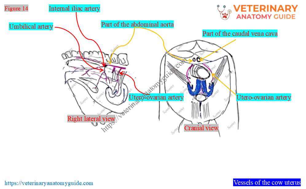
Cranial uterine artery: It is a branch of the utero-ovarian artery, a direct branch of the cow’s abdominal aorta. It supplies the cranial part of the cow’s uterine horn and anastomoses with the middle uterine artery.
Middle uterine artery: It is considered the main arterial supply for the cow’s uterus. However, it reaches the concave border of the horns through the broad ligament and divides into several branches.
Caudal uterine artery: A collateral branch of the internal iliac artery that supplies the ventral surface of the body and neck of the cow’s uterus.
Nerves: The cow’s uterus gets nerve innervation through the pelvic plexus.
FAQ’s on the cow’s uterus anatomy
The neck (cervix) of the cow’s uterus presents longitudinal folds and firm spiral plugs or rings. These structures give the neck a firm, dense appearance.
However, the neck wall is thicker than any other part of the uterus. So, while you palpate the neck of the fresh cow’s uterus, you will find it harder than other parts.
You will find a variation in the number of the intercornual ligaments between the cow and goat (Getty, 1977). In the cow, the uterus has dorsal and ventral intercornual ligaments, whereas the goat’s uterus has a dorsal intercornual ligament.
Conclusion
Thus, the cow uterus anatomy includes its three parts (horns, body, and neck) and their locations. Here, the two horns of the cow’s uterus present the most unique and oval caruncles internally.
Again, the body of the cow’s uterus is very small and has no unique features. However, the neck of the cow’s uterus has unique features such as the cervical canal, plugs/rings, and orifices.
References
- Monteiro, C. M. R.; et al. 2001. Comparative anatomy of uterus and oviducts of different cows. Braz. J. Vet. Res. Anim. Sci., 38(1):13-6.
- Ghosh, R. K.: Primary veterinary anatomy, 2nd Edn.Current Books International, Kolkata.
- Rowen D. Frandson, W. Lee Wilke, and A. Dee: Anatomy and physiology of farm animals, 4th edn.Wiley-Blackwell, USA.
- Monteiro CMR, et al. 2003. Study of histological characteristics of uterus and oviducts of cows. Braz. J. Vet. Res. Anim. Sci.: 40(1):45-54.
- Getty, R. Sission and Grossman’s The anatomy of the domestic animals, 5th Edn. W. B. Saunders and Co., Philadelphia (USA).
- Nickel, R., Schummer, A. and Seiferle, E.: The anatomy of domestic animals, 1st Edn. Verlag Paul Parey, Berlin.
- Neves, M.M., et al. (2002). Characteristics of the ovaries of the cows. Arq Bras De Medi Vet E Zoo, 54, 651-654.
- Hermanson et al., Miller and Evan’s Anatomy of the dog, 5th Edn. Elsevier.
- McLeod, W. N.: Bovine anatomy, 2nd Edn. Burgess Publishing Co., Minneapolis.
- Dyce, K. M., W. O. Sack, and CSG Wensing: Textbook of Veterinary Anatomy, Lea & Febiger. Philadelphia
Written by